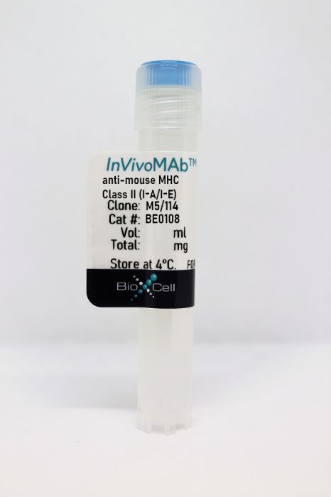
InVivoMAb anti-mouse MHC Class II (I-A/I-E)
Product Details
The M5/114 monoclonal antibody reacts with mouse MHC Class II haplotypes I-Ab, I-Ad, I-Aq, I-Ed, and I-Ek. The antibody does not react with I-Af, I-Ak, or I-As haplotypes. The M5/114 antibody is reported to inhibit I-A-restricted T cell responses.Specifications
| Isotype | Rat IgG2b |
|---|---|
| Recommended Isotype Control(s) | InVivoMAb rat IgG2b isotype control, anti-keyhole limpet hemocyanin |
| Recommended Dilution Buffer | InVivoPure pH 7.0 Dilution Buffer |
| Conjugation | This product is unconjugated. Conjugation is available via our Antibody Conjugation Services. |
| Immunogen | Activated C57BL/6 mouse spleen cells |
| Reported Applications |
in vivo MHC II blockade Functional assays Immunofluorescence Western blot Immunoprecipitation Flow cytometry |
| Formulation |
PBS, pH 7.0 Contains no stabilizers or preservatives |
| Endotoxin |
<2EU/mg (<0.002EU/μg) Determined by LAL gel clotting assay |
| Purity |
>95% Determined by SDS-PAGE |
| Sterility | 0.2 µm filtration |
| Production | Purified from cell culture supernatant in an animal-free facility |
| Purification | Protein G |
| RRID | AB_10949298 |
| Molecular Weight | 150 kDa |
| Storage | The antibody solution should be stored at the stock concentration at 4°C. Do not freeze. |
Recommended Products
-
Recommended Isotype Control(s)
InVivoMAb rat IgG2b isotype control, anti-keyhole limpet hemocyanin
-

Recommended Dilution Buffer
InVivoPure pH 7.0 Dilution Buffer
in vivo MHC II blockade
Kolodin, D., et al. (2015). "Antigen- and cytokine-driven accumulation of regulatory T cells in visceral adipose tissue of lean mice" Cell Metab 21(4): 543-557. PubMed
A unique population of Foxp3(+)CD4(+) regulatory T (Treg) cells, with a distinct transcriptome and antigen-receptor repertoire, resides in visceral adipose tissue (VAT) of lean individuals. These cells regulate local inflammation and both local and systemic metabolic indices. Here we focus on expansion of the VAT Treg compartment in aging lean mice-assessing these cells’ phenotypic conversion from conventional CD4(+) T cells, influx from lymphoid organs, and local population dynamics. Our findings establish that the VAT Treg compartment is seeded from thymocytes generated during the first weeks of life and expands beyond 10 weeks of age due to indolent proliferation, of certain clones in particular, coupled with enhanced survival. Accumulation of VAT Tregs depends on the antigen(s) presented by MHC class-II molecules and soluble mediators, notably interleukin(IL)-33. Addressing such factors therapeutically promises novel approaches for harnessing Tregs to stem the growing epidemic of obesity and consequent metabolic abnormalities.
Functional Assays
Kreiter, S., et al. (2015). "Mutant MHC class II epitopes drive therapeutic immune responses to cancer" Nature 520(7549): 692-696. PubMed
Tumour-specific mutations are ideal targets for cancer immunotherapy as they lack expression in healthy tissues and can potentially be recognized as neo-antigens by the mature T-cell repertoire. Their systematic targeting by vaccine approaches, however, has been hampered by the fact that every patient’s tumour possesses a unique set of mutations (‘the mutanome’) that must first be identified. Recently, we proposed a personalized immunotherapy approach to target the full spectrum of a patient’s individual tumour-specific mutations. Here we show in three independent murine tumour models that a considerable fraction of non-synonymous cancer mutations is immunogenic and that, unexpectedly, the majority of the immunogenic mutanome is recognized by CD4(+) T cells. Vaccination with such CD4(+) immunogenic mutations confers strong antitumour activity. Encouraged by these findings, we established a process by which mutations identified by exome sequencing could be selected as vaccine targets solely through bioinformatic prioritization on the basis of their expression levels and major histocompatibility complex (MHC) class II-binding capacity for rapid production as synthetic poly-neo-epitope messenger RNA vaccines. We show that vaccination with such polytope mRNA vaccines induces potent tumour control and complete rejection of established aggressively growing tumours in mice. Moreover, we demonstrate that CD4(+) T cell neo-epitope vaccination reshapes the tumour microenvironment and induces cytotoxic T lymphocyte responses against an independent immunodominant antigen in mice, indicating orchestration of antigen spread. Finally, we demonstrate an abundance of mutations predicted to bind to MHC class II in human cancers as well by employing the same predictive algorithm on corresponding human cancer types. Thus, the tailored immunotherapy approach introduced here may be regarded as a universally applicable blueprint for comprehensive exploitation of the substantial neo-epitope target repertoire of cancers, enabling the effective targeting of every patient’s tumour with vaccines produced ‘just in time’.
in vivo MHC II blockade, Flow Cytometry
McKinstry, K. K., et al. (2014). "Effector CD4 T-cell transition to memory requires late cognate interactions that induce autocrine IL-2" Nat Commun 5: 5377. PubMed
It is unclear how CD4 T-cell memory formation is regulated following pathogen challenge, and when critical mechanisms act to determine effector T-cell fate. Here, we report that following influenza infection most effectors require signals from major histocompatibility complex class II molecules and CD70 during a late window well after initial priming to become memory. During this timeframe, effector cells must produce IL-2 or be exposed to high levels of paracrine or exogenously added IL-2 to survive an otherwise rapid default contraction phase. Late IL-2 promotes survival through acute downregulation of apoptotic pathways in effector T cells and by permanently upregulating their IL-7 receptor expression, enabling IL-7 to sustain them as memory T cells. This new paradigm defines a late checkpoint during the effector phase at which cognate interactions direct CD4 T-cell memory generation.
in vivo MHC II blockade, Flow Cytometry
Fu, H., et al. (2014). "Self-recognition of the endothelium enables regulatory T-cell trafficking and defines the kinetics of immune regulation" Nat Commun 5: 3436. PubMed
Localization of CD4(+)CD25(+)Foxp3(+) regulatory T (Treg) cells to lymphoid and non-lymphoid tissue is instrumental for the effective control of immune responses. Compared with conventional T cells, Treg cells constitute a minute fraction of the T-cell repertoire. Despite this numeric disadvantage, Tregs efficiently migrate to sites of immune responses reaching an optimal number for the regulation of T effector (Teff) cells. The array and levels of adhesion and chemokine receptor expression by Tregs do not explain their powerful migratory capacity. Here we show that recognition of self-antigens expressed by endothelial cells in target tissue is instrumental for efficient Treg recruitment in vivo. This event relies upon IFN-gamma-mediated induction of MHC-class-II molecule expression by the endothelium and requires optimal PI3K p110delta activation by the T-cell receptor. We also show that, once in the tissue, Tregs inhibit Teff recruitment, further enabling a Teff:Treg ratio optimal for regulation.
Functional Assays
Bar, E., et al. (2012). "A novel Th cell epitope of Candida albicans mediates protection from fungal infection" J Immunol 188(11): 5636-5643. PubMed
Fungal pathogens are a frequent cause of opportunistic infections. They live as commensals in healthy individuals but can cause disease when the immune status of the host is altered. T lymphocytes play a critical role in pathogen control. However, specific Ags determining the activation and function of antifungal T cells remain largely unknown. By using an immunoproteomic approach, we have identified for the first time, to our knowledge, a natural T cell epitope from Candida albicans. Isolation and sequencing of MHC class II-bound ligands from infected dendritic cells revealed a peptide that was recognized by a major population of all Candida-specific Th cells isolated from infected mice. Importantly, human Th cells also responded to stimulation with the peptide in an HLA-dependent manner but without restriction to any particular HLA class II allele. Immunization of mice with the peptide resulted in a population of epitope-specific Th cells that reacted not only with C. albicans but also with other clinically highly relevant species of Candida including the distantly related Candida glabrata. The extent of the reaction to different Candida species correlated with their degree of phylogenetic relationship to C. albicans. Finally, we show that the newly identified peptide acts as an efficient vaccine when used in combination with an adjuvant inducing IL-17A secretion from peptide-specific T cells. Immunized mice were protected from fatal candidiasis. Together, these results uncover a new immune determinant of the host response against Candida ssp. that could be exploited for the development of antifungal vaccines and immunotherapies.
Flow Cytometry
Rockett, B. D., et al. (2010). "n-3 PUFA improves fatty acid composition, prevents palmitate-induced apoptosis, and differentially modifies B cell cytokine secretion in vitro and ex vivo" J Lipid Res 51(6): 1284-1297. PubMed
n-3 polyunsaturated fatty acids (PUFAs) modify T-cell activation, in part by remodeling lipid composition; however, the relationship between n-3 PUFA and B-cell activation is unknown. Here we tested this relationship in vitro and ex vivo by measuring upregulation of B-cell surface molecules, the percentage of cells activated, and cytokine secreted in response to lipopolysaccharide (LPS) activation. In vitro, eicosapentaenoic acid (EPA) and docosahexaenoic acid (DHA) improved the membrane n-6/n-3 PUFA ratio, and DHA lowered interleukin (IL)-6 secretion; overall, n-3 PUFAs did not suppress B-cell activation compared with BSA, oleate, or elaidate treatment. Palmitate treatment suppressed the percentage of B cells activated through lipoapoptosis, which was differentially prevented by cosupplementing cells with MUFAs and PUFAs. Ex vivo, we tested the hypothesis with mice fed a control or high-fat saturated, hydrogenated, MUFA or n-3 PUFA diets. n-3 PUFAs had no effect on the percentage of B cells activated. Unexpectedly, the n-3 PUFA diet increased B-cell CD69 surface expression, IL-6 and IFNgamma secretion, and it significantly increased body weight gain. Overall, we propose that changes in lipid composition with n-3 PUFA and suppression of lymphocyte activation is not universal. The study highlights that high-fat n-3 PUFA diets can promote pro-inflammatory responses, at least from one cell type.
in vivo MHC II blockade
Quezada, S. A., et al. (2010). "Tumor-reactive CD4(+) T cells develop cytotoxic activity and eradicate large established melanoma after transfer into lymphopenic hosts" J Exp Med 207(3): 637-650. PubMed
Adoptive transfer of large numbers of tumor-reactive CD8(+) cytotoxic T lymphocytes (CTLs) expanded and differentiated in vitro has shown promising clinical activity against cancer. However, such protocols are complicated by extensive ex vivo manipulations of tumor-reactive cells and have largely focused on CD8(+) CTLs, with much less emphasis on the role and contribution of CD4(+) T cells. Using a mouse model of advanced melanoma, we found that transfer of small numbers of naive tumor-reactive CD4(+) T cells into lymphopenic recipients induces substantial T cell expansion, differentiation, and regression of large established tumors without the need for in vitro manipulation. Surprisingly, CD4(+) T cells developed cytotoxic activity, and tumor rejection was dependent on class II-restricted recognition of tumors by tumor-reactive CD4(+) T cells. Furthermore, blockade of the coinhibitory receptor CTL-associated antigen 4 (CTLA-4) on the transferred CD4(+) T cells resulted in greater expansion of effector T cells, diminished accumulation of tumor-reactive regulatory T cells, and superior antitumor activity capable of inducing regression of spontaneous mouse melanoma. These findings suggest a novel potential therapeutic role for cytotoxic CD4(+) T cells and CTLA-4 blockade in cancer immunotherapy, and demonstrate the potential advantages of differentiating tumor-reactive CD4(+) cells in vivo over current protocols favoring in vitro expansion and differentiation.
Western Blot, Immunoprecipitation
Li, C., et al. (2002). "Cooperative interaction of Ig(alpha) and Ig(beta) of the BCR regulates the kinetics and specificity of antigen targeting" Int Immunol 14(10): 1179-1191. PubMed
Following the binding of antigens, the BCR transduces signals and internalizes antigens for processing and presentation, both of which are required for initiating an effective antibody response. The BCR, consisting of membrane Ig and Ig(alpha)/Ig(beta) heterodimer, facilitates antigen processing by accelerating antigen targeting to the processing compartment. Previous reports showed that Ig(alpha) or Ig(beta) alone is competent for internalizing antigens. However, both Ig(alpha) and Ig(beta) are required for BCR-enhanced antigen presentation. Using chimeric proteins containing the extracellular and transmembrane domains of human platelet-derived growth factor receptor fused with the cytoplasmic domain of Ig(alpha) or Ig(beta), we studied the roles of the cytoplasmic tails of Ig(alpha) and Ig(beta) in BCR-mediated antigen transport. The Ig(beta) chimera rapidly moves through the endocytic pathway to lysosomes, while the Ig(alpha) chimera slows down this movement. The Ig(alpha), but not the Ig(beta) chimera, is required for an increase in the turnover rate of the chimeras in response to stimulation. Only when Ig(alpha) and Ig(beta) chimeras are co-expressed do the chimeras rapidly and specifically target antigens to the processing compartment. These findings suggest that Ig(alpha) and Ig(beta) play distinct roles in BCR trafficking, and the cooperative interaction of Ig(alpha) and Ig(beta) controls and regulates the kinetics and specificity of antigen targeting.
Immunofluorescence, Immunoprecipitation
Pierre, P., et al. (1997). "Developmental regulation of MHC class II transport in mouse dendritic cells" Nature 388(6644): 787-792. PubMed
Dendritic cells (DCs) have the unique capacity to initiate primary and secondary immune responses. They acquire antigens in peripheral tissues and migrate to lymphoid organs where they present processed peptides to T cells. DCs must therefore exist in distinct functional states, an idea that is supported by observations that they downregulate endocytosis and upregulate surface molecules of the class II major histocompatibility complex (MHC) upon maturation. Here we investigate the features of DC maturation by reconstituting the terminal differentiation of mouse DCs in vitro and in situ. We find that early DCs, corresponding to those found in peripheral tissues, exhibit a phenotype in which most class II molecules are intracellular and localized to lysosomes. Upon maturation, these cells give rise to a new intermediate phenotype in which intracellular class II molecules are found in peripheral non-lysosomal vesicles, similar to the specialized CIIV population seen in B cells. The intermediate cells then differentiate into late DCs which express almost all of their class II molecules on the plasma membrane. These variations in class II compartmentalization are accompanied by dramatic alterations in the intracellular transport of the new class II molecules and in antigen presentation. We found that although early DCs could not present antigen immediately after uptake, efficient presentation of the previously internalized antigen occurred after maturation, 24-48 hours later. By regulating class II transport and compartmentalization, DCs are able to delay antigen display, a property crucial to their role in immune surveillance.
- Mus musculus (House mouse),
- Immunology and Microbiology
Attenuated IL-2 muteins leverage the TCR signal to enhance regulatory T cell homeostasis and response in vivo.
In Frontiers in Immunology on 9 October 2023 by Ma, S., So, M., et al.
PubMed
Interleukin-2 (IL-2), along with T-cell receptor (TCR) signaling, are required to control regulatory T cell (Treg) homeostasis and function in vivo. Due to the heightened sensitivity to IL-2, Tregs retain the ability to respond to low-dose or attenuated forms of IL-2, as currently being developed for clinical use to treat inflammatory diseases. While attenuated IL-2 increases Treg selectivity, the question remains as to whether a weakened IL-2 signal sufficiently enhances Treg suppressive function(s) toward disease modification. To understand this question, we characterized the in vivo activity and transcriptomic profiles of two different attenuated IL-2 muteins in comparison with wildtype (WT) IL-2. Our study showed that, in addition to favoring Tregs, the attenuated muteins induced disproportionately robust effects on Treg activation and conversion to effector Treg (eTreg) phenotype. Our data furthermore suggested that Tregs activated by attenuated IL-2 muteins showed reduced dependence on TCR signal, at least in part due to the enhanced ability of IL-2 muteins to amplify the TCR signal in vivo. These results point to a new paradigm wherein IL-2 influences Tregs' sensitivity to antigenic signal, and that the combination effect may be leveraged for therapeutic use of attenuated IL-2 muteins. Copyright © 2023 Ma, So, Ghelani, Srivas, Sahoo, Hall, Liu, Wu, Yu, Lu, Song, Cariaga, Soto, Zhou, Li, Chaudhry, Luo and Sohn.
- Immunology and Microbiology
The gut microbiota promotes distal tissue regeneration via RORγ+ regulatory T cell emissaries.
In Immunity on 11 April 2023 by Hanna, B. S., Wang, G., et al.
PubMed
Specific microbial signals induce the differentiation of a distinct pool of RORγ+ regulatory T (Treg) cells crucial for intestinal homeostasis. We discovered highly analogous populations of microbiota-dependent Treg cells that promoted tissue regeneration at extra-gut sites, notably acutely injured skeletal muscle and fatty liver. Inflammatory meditators elicited by tissue damage combined with MHC-class-II-dependent T cell activation to drive the accumulation of gut-derived RORγ+ Treg cells in injured muscle, wherein they regulated the dynamics and tenor of early inflammation and helped balance the proliferation vs. differentiation of local stem cells. Reining in IL-17A-producing T cells was a major mechanism underlying the rheostatic functions of RORγ+ Treg cells in compromised tissues. Our findings highlight the importance of gut-trained Treg cell emissaries in controlling the response to sterile injury of non-mucosal tissues. Copyright © 2023 Elsevier Inc. All rights reserved.
- FC/FACS,
- Mus musculus (House mouse),
- Immunology and Microbiology
Group 2 innate lymphoid cells mediate the activation of CD4+ T cells and aggravate Th1/Th2 imbalance via MHC II molecules during respiratory syncytial virus infection.
In International Immunopharmacology on 1 December 2022 by Kang, L., Wang, S., et al.
PubMed
Respiratory syncytial virus (RSV) infection induces the activation of CD4+ T cells. However, the underlying mechanism of CD4+T-cell activation induced by RSV infection is not fully understood. In the present study, we found that depletion of CD4+ T cells can obviously reduce airway inflammation caused by RSV infection. Meanwhile, adoptive transfer of group 2 innate lymphocytes (ILC2s) significantly enhanced the number of CD4+ T cells and promoted their differentiation to Th2 in lung. In fact, RSV infection increased the expression of major histocompatibility complex-II (MHC II) molecules on the surface of pulmonary ILC2s. In vitro coculture experiments showed that ILC2s may act as promoters to promote the expansion and differentiation of RSV-infected CD4+ T cells. However, blocking the interaction between CD4+ T cells and ILC2s with anti-MHC-II mAbs significantly reduced CD4+T-cell expansion. These results suggest that pulmonary ILC2s may function as antigen-presenting cells to induce the activation of CD4+ T cells through the MHC II pathway during RSV infection. Copyright © 2022 Elsevier B.V. All rights reserved.
- Cancer Research
Agonistic anti-CD40 converts Tregs into Type 1 effectors within the tumor micro-environment
Preprint on BioRxiv : the Preprint Server for Biology on 20 October 2022 by Maltez, V., Arora, C., et al.
PubMed
Multiple cell types, molecules, and processes contribute to inhibition of anti-tumor effector responses, often frustrating effective immunotherapy. Among these, Foxp3+ CD4+ cells (Tregs) are well-recognized to play an immunosuppressive role in the tumor microenvironment. The first clinically successful checkpoint inhibitor, anti-CTLA-4 antibody, may deplete Tregs at least in part by antibody-dependent cellular cytotoxicity (ADCC), but this effect is unreliable in mice, including in a genetically engineered mouse model of pancreatic ductal adenocarcinoma (PDAC). In contrast, agonistic CD40 antibody, which serves as an effective therapy, is associated with notable Treg disappearance in the PDAC model. The mechanism of CD40-mediated Treg loss is poorly understood, as Tregs are CD40-negative. Here we have explored the mechanistic basis for the loss of Foxp3 T cells upon anti-CD40 treatment and find, using tissue-level multiplex immunostaining and orthogonal dissociated cell analyses, that Tregs are not depleted but converted into interferon- γ (IFN- γ ) producing, Type I CD4+ T effector cells. This process depends on IL-12 and IFN- γ signaling evoked by action of the anti-CD40 antibody on dendritic cells (DCs), especially BATF3-dependent cDC1s. These findings provide insight into a previously unappreciated mechanism of CD40 agonism as a potent anti-tumor intervention that promotes the re-programming of Tregs into tumor-reactive CD4+ effector T cells, markedly augmenting the anti-tumor response.
- Cancer Research,
- Immunology and Microbiology
Cancer-specific T helper shared and neo-epitopes uncovered by expression of the MHC class II master regulator CIITA.
In Cell Reports on 11 October 2022 by Hos, B. J., Tondini, E., et al.
PubMed
We report an approach to identify tumor-specific CD4+ T cell neo-epitopes of both mouse and human cancer cells by analysis of major histocompatibility complex (MHC) class II-eluted natural peptides. MHC class II-presented peptide sequences are identified by introducing the MHC class II transactivator (CIITA) in tumor cells that were originally MHC class II negative. CIITA expression facilitates cell-surface expression of MHC class II molecules and the appropriate peptide-loading machinery. Peptide elution of purified MHC class II molecules and subsequent mass spectrometry reveals oncoviral- and neo-epitopes as well as shared epitopes. Immunological relevance of these epitopes is shown by natural presentation by dendritic cells and immunogenicity. Synthetic peptide vaccination induced functional CD4+ T cell responses, which helped tumor control in vivo. Thus, this CIITA transfection approach aids to identify relevant T helper epitopes presented by any MHC class II allele that would be otherwise very difficult to predict and reveals important targets for cancer immunotherapy. Copyright © 2022 The Authors. Published by Elsevier Inc. All rights reserved.
- Mus musculus (House mouse),
- Immunology and Microbiology
Tfh-cell-derived interleukin 21 sustains effector CD8+ T cell responses during chronic viral infection.
In Immunity on 8 March 2022 by Zander, R., Kasmani, M. Y., et al.
PubMed
CD4+ T cell-derived interleukin 21 (IL-21) sustains CD8+ T cell responses during chronic viral infection, but the helper subset that confers this protection remains unclear. Here, we applied scRNA and ATAC-seq approaches to determine the heterogeneity of IL-21+CD4+ T cells during LCMV clone 13 infection. CD4+ T cells were comprised of three transcriptionally and epigenetically distinct populations: Cxcr6+ Th1 cells, Cxcr5+ Tfh cells, and a previously unrecognized Slamf6+ memory-like (Tml) subset. T cell differentiation was specifically redirected toward the Tml subset during chronic, but not acute, LCMV infection. Although this subset displayed an enhanced capacity to accumulate and some developmental plasticity, it remained largely quiescent, which may hinder its helper potential. Conversely, mixed bone marrow chimera experiments revealed that Tfh cell-derived IL-21 was critical to sustain CD8+ T cell responses and viral control. Thus, strategies that bolster IL-21+Tfh cell responses may prove effective in enhancing CD8+ T cell-mediated immunity. Copyright © 2022 Elsevier Inc. All rights reserved.
- Cancer Research,
- Immunology and Microbiology,
- Cell Culture,
- Mus musculus (House mouse)
Lung tumor MHCII immunity depends on in situ antigen presentation by fibroblasts.
In The Journal of Experimental Medicine on 7 February 2022 by Kerdidani, D., Aerakis, E., et al.
PubMed
A key unknown of the functional space in tumor immunity is whether CD4 T cells depend on intratumoral MHCII cancer antigen recognition. MHCII-expressing, antigen-presenting cancer-associated fibroblasts (apCAFs) have been found in breast and pancreatic tumors and are considered to be immunosuppressive. This analysis shows that antigen-presenting fibroblasts are frequent in human lung non-small cell carcinomas, where they seem to actively promote rather than suppress MHCII immunity. Lung apCAFs directly activated the TCRs of effector CD4 T cells and at the same time produced C1q, which acted on T cell C1qbp to rescue them from apoptosis. Fibroblast-specific MHCII or C1q deletion impaired CD4 T cell immunity and accelerated tumor growth, while inducing C1qbp in adoptively transferred CD4 T cells expanded their numbers and reduced tumors. Collectively, we have characterized in the lungs a subset of antigen-presenting fibroblasts with tumor-suppressive properties and propose that cancer immunotherapies might be strongly dependent on in situ MHCII antigen presentation. © 2022 Kerdidani et al.
- FC/FACS,
- Mus musculus (House mouse),
- Immunology and Microbiology
Group 2 innate lymphoid cells mediate the activation of CD4+ T cells and aggravate Th1/Th2 imbalance via MHC II molecules during respiratory syncytial virus infection
Preprint on Research Square on 25 October 2021 by Kang, L., Wang, S., et al.
PubMed
h4>Background: /h4> Respiratory syncytial virus (RSV), as an important worldwide pediatric respiratory pathogen, can cause pneumonia and bronchiolitis in infants and young children. CD4 + T cells play a crucial role in RSV-induced airway inflammation. However, the underlying mechanism of CD4 + T cell activation during RSV infection is not fully understood. Methods The proliferation and activation of CD4 + T cells in the lungs of RSV infected BALB/c mice were measured by flow cytometry. RSV-induced airway inflammation was determined by HE staining and the inflammatory cells in bronchoalveolar lavage fluids (BALF) were counted. The protein levels of IL-4, IL-5, IL-13, and IFN-γ in the supernatant of BALF were detected by ELISA. The mRNA levels of IL-4, IL-5, IL-13, and IFN-γ in the lung homogenates were analyzed by real-time PCR. The expression of major histocompatibility complex II (MHCII), CD80 and CD86 on pulmonary ILC2s were measured by flow cytometry and real-time PCR. Results RSV infection may promote the differentiation and activation of CD4 + Th2 cells, resulting in imbalance of Th1/Th2 in the lung tissues. Depletion of CD4 + T cells can obviously reduce airway inflammation caused by RSV infection. Adoptive transfer of pulmonary ILC2s significantly enhanced the number of pulmonary CD4 + T cells and promoted their differentiation to Th2. In fact, RSV infection increased the expression of MHC II and B7 molecules on the surface of pulmonary ILC2s. In vitro co-culture experiments showed that ILC2s may act as a promoter to promote the expansion and differentiation of RSV-infected naïve CD4 + T cells. However, blocking the interaction between ILC2s and CD4 + T cells with anti-MHC II mAbs significantly reduced CD4 + T cell expansion and increased the Th1/Th2 ratio of CD4 + T cells. Conclusions Our data suggest that pulmonary ILC2s may function as antigen-presenting cells to induce the activation of CD4 + T cells through MHC II pathway during RSV infection.
CXCL10+ peripheral activation niches couple preferred sites of Th1 entry with optimal APC encounter.
In Cell Reports on 10 August 2021 by Prizant, H., Patil, N., et al.
PubMed
Correct positioning of T cells within infected tissues is critical for T cell activation and pathogen control. Upon tissue entry, effector T cells must efficiently locate antigen-presenting cells (APC) for peripheral activation. We reveal that tissue entry and initial peripheral activation of Th1 effector T cells are tightly linked to perivascular positioning of chemokine-expressing APCs. Dermal inflammation induces tissue-wide de novo generation of discrete perivascular CXCL10+ cell clusters, enriched for CD11c+MHC-II+ monocyte-derived dendritic cells. These chemokine clusters are "hotspots" for both Th1 extravasation and activation in the inflamed skin. CXCR3-dependent Th1 localization to the cluster micro-environment prolongs T-APC interactions and boosts function. Both the frequency and range of these clusters are enhanced via a T helper 1 (Th1)-intrinsic, interferon-gamma (IFNγ)-dependent positive-feedback loop. Thus, the perivascular CXCL10+ clusters act as initial peripheral activation niches, optimizing controlled activation broadly throughout the tissue by coupling Th1 tissue entry with enhanced opportunities for Th1-APC encounter. Copyright © 2021. Published by Elsevier Inc.
- Immunology and Microbiology
Targeting Treg cells with GITR activation alleviates resistance to immunotherapy in murine glioblastomas.
In Nature Communications on 11 May 2021 by Amoozgar, Z., Kloepper, J., et al.
PubMed
Immune checkpoint blockers (ICBs) have failed in all phase III glioblastoma (GBM) trials. Here, we show that regulatory T (Treg) cells play a key role in GBM resistance to ICBs in experimental gliomas. Targeting glucocorticoid-induced TNFR-related receptor (GITR) in Treg cells using an agonistic antibody (αGITR) promotes CD4 Treg cell differentiation into CD4 effector T cells, alleviates Treg cell-mediated suppression of anti-tumor immune response, and induces potent anti-tumor effector cells in GBM. The reprogrammed GBM-infiltrating Treg cells express genes associated with a Th1 response signature, produce IFNγ, and acquire cytotoxic activity against GBM tumor cells while losing their suppressive function. αGITR and αPD1 antibodies increase survival benefit in three experimental GBM models, with a fraction of cohorts exhibiting complete tumor eradication and immune memory upon tumor re-challenge. Moreover, αGITR and αPD1 synergize with the standard of care treatment for newly-diagnosed GBM, enhancing the cure rates in these GBM models.
- Chemistry
Application of a Highly Selective Cathepsin S Two-step Activity-Based Probe in Multicolor Bio-Orthogonal Correlative Light-Electron Microscopy.
In Frontiers in Chemistry on 2 March 2021 by van Dalen, F. J., Bakkum, T., et al.
PubMed
Cathepsin S is a lysosomal cysteine protease highly expressed in immune cells such as dendritic cells, B cells and macrophages. Its functions include extracellular matrix breakdown and cleavage of cell adhesion molecules to facilitate immune cell motility, as well as cleavage of the invariant chain during maturation of major histocompatibility complex II. The identification of these diverse specific functions has brought the challenge of delineating cathepsin S activity with great spatial precision, relative to related enzymes and substrates. Here, the development of a potent and highly selective two-step activity-based probe for cathepsin S and the application in multicolor bio-orthogonal correlative light-electron microscopy is presented. LHVS, which has been reported as a selective inhibitor of cathepsin S with nanomolar potency, formed the basis for our probe design. However, in competitive activity-based protein profiling experiments LHVS showed significant cross-reactivity toward Cat L. Introduction of an azide group in the P2 position expanded the selectivity window for cathepsin S, but rendered the probe undetectable, as demonstrated in bio-orthogonal competitive activity-based protein profiling. Incorporation of an additional azide handle for click chemistry on the solvent-exposed P1 position allowed for selective labeling of cathepsin S. This highlights the influence of click handle positioning on probe efficacy. This probe was utilized in multicolor bio-orthogonal confocal and correlative light-electron microscopy to investigate the localization of cathepsin S activity at an ultrastructural level in bone marrow-derived dendritic cells. The tools developed in this study will aid the characterization of the variety of functions of cathepsin S throughout biology. Copyright © 2021 van Dalen, Bakkum, van Leeuwen, Groenewold, Deu, Koster, van Kasteren and Verdoes.
- Block,
- Mus musculus (House mouse),
- Cancer Research,
- Immunology and Microbiology
Radiotherapy-exposed CD8+ and CD4+ neoantigens enhance tumor control.
In The Journal of Clinical Investigation on 1 March 2021 by Lhuillier, C., Rudqvist, N. P., et al.
PubMed
Neoantigens generated by somatic nonsynonymous mutations are key targets of tumor-specific T cells, but only a small number of mutations predicted to be immunogenic are presented by MHC molecules on cancer cells. Vaccination studies in mice and patients have shown that the majority of neoepitopes that elicit T cell responses fail to induce significant antitumor activity, for incompletely understood reasons. We report that radiotherapy upregulates the expression of genes containing immunogenic mutations in a poorly immunogenic mouse model of triple-negative breast cancer. Vaccination with neoepitopes encoded by these genes elicited CD8+ and CD4+ T cells that, whereas ineffective in preventing tumor growth, improved the therapeutic efficacy of radiotherapy. Mechanistically, neoantigen-specific CD8+ T cells preferentially killed irradiated tumor cells. Neoantigen-specific CD4+ T cells were required for the therapeutic efficacy of vaccination and acted by producing Th1 cytokines, killing irradiated tumor cells, and promoting epitope spread. Such a cytotoxic activity relied on the ability of radiation to upregulate class II MHC molecules as well as the death receptors FAS/CD95 and DR5 on the surface of tumor cells. These results provide proof-of-principle evidence that radiotherapy works in concert with neoantigen vaccination to improve tumor control.
- In Vivo,
- Mus musculus (House mouse),
- Immunology and Microbiology
A conjoined universal helper epitope can unveil antitumor effects of a neoantigen vaccine targeting an MHC class I-restricted neoepitope.
In NPJ Vaccines on 18 January 2021 by Swartz, A. M., Congdon, K. L., et al.
PubMed
Personalized cancer vaccines targeting neoantigens arising from somatic missense mutations are currently being evaluated for the treatment of various cancers due to their potential to elicit a multivalent, tumor-specific immune response. Several cancers express a low number of neoantigens; in these cases, ensuring the immunotherapeutic potential of each neoantigen-derived epitope (neoepitope) is crucial. In this study, we discovered that therapeutic vaccines targeting immunodominant major histocompatibility complex (MHC) I-restricted neoepitopes require a conjoined helper epitope in order to induce a cytotoxic, neoepitope-specific CD8+ T-cell response. Furthermore, we show that the universally immunogenic helper epitope P30 can fulfill this requisite helper function. Remarkably, conjoined P30 was able to unveil immune and antitumor responses to subdominant MHC I-restricted neoepitopes that were, otherwise, poorly immunogenic. Together, these data provide key insights into effective neoantigen vaccine design and demonstrate a translatable strategy using a universal helper epitope that can improve therapeutic responses to MHC I-restricted neoepitopes.
- Immunology and Microbiology
Immunopeptidomics for Dummies: Detailed Experimental Protocols and Rapid, User-Friendly Visualization of MHC I and II Ligand Datasets with MhcVizPipe
Preprint on BioRxiv : the Preprint Server for Biology on 3 November 2020 by Kovalchik, K. A., Wessling, L., et al.
PubMed
h4>ABSTRACT/h4> Immunopeptidomics refers to the science of investigating the composition and dynamics of peptides presented by major histocompatibility complex (MHC) class I and class II molecules using mass spectrometry (MS). Here, we aim to provide a technical report to any non-expert in the field wishing to establish and/or optimize an immunopeptidomic workflow with relatively limited computational knowledge and resources. To this end, we thoroughly describe step-by-step instructions to isolate MHC class I and II-associated peptides from various biological sources, including mouse and human biospecimens. Most notably, we created MhcVizPipe (MVP) ( https://github.com/CaronLab/MhcVizPipe ), a new and easy-to-use open-source software tool to rapidly assess the quality and the specific enrichment of immunopeptidomic datasets upon the establishment of new workflows. In fact, MVP enables intuitive visualization of multiple immunopeptidomic datasets upon testing sample preparation protocols and new antibodies for the isolation of MHC class I and II peptides. In addition, MVP enables the identification of unexpected binding motifs and facilitates the analysis of non-canonical MHC peptides. We anticipate that the experimental and bioinformatic resources provided herein will represent a great starting point for any non-expert and will therefore foster the accessibility and expansion of the field to ultimately boost its maturity and impact.
- Immunology and Microbiology
Regulatory T Cell Transmigration and Intravascular Migration Undergo Mechanistically Distinct Regulation at Different Phases of the Inflammatory Response.
In The Journal of Immunology on 1 December 2019 by Snelgrove, S. L., Abeynaike, L. D., et al.
PubMed
Regulatory T cells (Tregs) play important roles in limiting inflammatory responses in the periphery. During these responses, Treg abundance in affected organs increases and interfering with their recruitment results in exacerbation of inflammation. However, the mechanisms whereby Tregs enter the skin remain poorly understood. The aim of this study was to use intravital microscopy to investigate adhesion and transmigration of Tregs in the dermal microvasculature in a two-challenge model of contact sensitivity. Using intravital confocal microscopy of Foxp3-GFP mice, we visualized endogenous Tregs and assessed their interactions in the dermal microvasculature. Four hours after hapten challenge, Tregs underwent adhesion with ∼25% of these cells proceeding to transmigration, a process dependent on CCR4. At 24 h, Tregs adhered but no longer underwent transmigration, instead remaining in prolonged contact with the endothelium, migrating over the endothelial surface. Four hours after a second challenge, Treg transmigration was restored, although in this case transmigration was CCR4 independent, instead involving the CCR6/CCL20 pathway. Notably, at 24 h but not 4 h after challenge, endothelial cells expressed MHC class II (MHC II). Moreover, at this time of peak MHC II expression, inhibition of MHC II reduced Treg adhesion, demonstrating an unexpected role for MHC II in Treg attachment to the endothelium. Together these data show that Treg adhesion and transmigration can be driven by different molecular mechanisms at different stages of an Ag-driven inflammatory response. In addition, Tregs can undergo prolonged migration on the inflamed endothelium. Copyright © 2019 by The American Association of Immunologists, Inc.
- In Vivo,
- Block,
- Mus musculus (House mouse),
- Immunology and Microbiology
The Chemoattractant Receptor Ebi2 Drives Intranodal Naive CD4+ T Cell Peripheralization to Promote Effective Adaptive Immunity.
In Immunity on 21 May 2019 by Baptista, A. P., Gola, A., et al.
PubMed
Lymph nodes (LNs) play critical roles in adaptive immunity by concentrating in one location the antigens, antigen-presenting cells, and antigen-responsive lymphocytes involved in such responses. Recent studies have revealed nonrandom localization of innate and adaptive immune cells within these organs, suggesting that microanatomical positioning optimizes responses involving sparse cooperating cells. Here, we report that the peripheral localization of LN cDC2 dendritic cells specialized for MHC-II antigen presentation is matched by a similarly biased paracortical distribution of CD4+ T cells directed by the chemoattractant receptor Ebi2. In the absence of Ebi2, CD4+ T cells lose their location bias and are delayed in antigen recognition, proliferative expansion, differentiation, direct effector activity, and provision of help for CD8+ T cell-mediated memory responses, limiting host defense and vaccine responses. These findings demonstrate evolutionary selection for distinct niches within the LN that promote cellular responses, emphasizing the critical link between fine-grained tissue organization and host defense.Published by Elsevier Inc.
- Genetics
Regulation of invariant NKT cell development and function by a 0.14 Mbp locus on chromosome 1: a possible role for Fcgr3.
In Genes and Immunity on 1 April 2019 by DeVault, V. L., Malagic, M., et al.
PubMed
Invariant NKT (iNKT) cells are tissue-resident innate-like T cells critical to the host immune response. We previously identified a 6.6 Mbp region on chromosome 1 as a major regulator of iNKT cell number and function in C57BL/6 and 129X1/SvJ mice. Here, we fine-mapped this locus by assessing the iNKT cell response to alpha-galactosylceramide (αGalCer) in a series of B6.129 congenic lines. This analysis revealed the presence of at least two genetic elements that regulate iNKT cell cytokine production in response to αGalCer. While one of these genetic elements mapped to the B6.129c6 interval containing Slam genes, the dominant regulator in this region mapped to the 0.14 Mbp B6.129c3 interval. In addition, we found that numbers of thymic iNKT cells and DP thymocytes were significantly lower in B6.129c3 mice, indicating that this interval also regulates iNKT cell development. Candidate gene analysis revealed a fivefold increase in Fcgr3 expression in B6.129c3 iNKT cells, and we observed increased expression of FcγR3 protein on B6.129c3 iNKT cells, NK cells, and neutrophils. These data identify the B6.129c3 interval as a novel locus regulating the response of iNKT cells to glycosphingolipid, revealing a link between this phenotype and a polymorphism that regulates Fcgr3 expression.
- Biochemistry and Molecular biology,
- Cell Biology
A timer for analyzing temporally dynamic changes in transcription during differentiation in vivo.
In The Journal of Cell Biology on 6 August 2018 by Bending, D., Prieto Martín, P., et al.
PubMed
Understanding the mechanisms of cellular differentiation is challenging because differentiation is initiated by signaling pathways that drive temporally dynamic processes, which are difficult to analyze in vivo. We establish a new tool, Timer of cell kinetics and activity (Tocky; or toki [time in Japanese]). Tocky uses the fluorescent Timer protein, which spontaneously shifts its emission spectrum from blue to red, in combination with computer algorithms to reveal the dynamics of differentiation in vivo. Using a transcriptional target of T cell receptor (TCR) signaling, we establish Nr4a3-Tocky to follow downstream effects of TCR signaling. Nr4a3-Tocky reveals the temporal sequence of events during regulatory T cell (Treg) differentiation and shows that persistent TCR signals occur during Treg generation. Remarkably, antigen-specific T cells at the site of autoimmune inflammation also show persistent TCR signaling. In addition, by generating Foxp3-Tocky, we reveal the in vivo dynamics of demethylation of the Foxp3 gene. Thus, Tocky is a tool for cell biologists to address previously inaccessible questions by directly revealing dynamic processes in vivo. © 2018 Bending et al.
- Immunology and Microbiology
Staphylococcus aureus Lipoprotein Induces Skin Inflammation, Accompanied with IFN-γ-Producing T Cell Accumulation through Dermal Dendritic Cells.
In Pathogens on 29 July 2018 by Saito, S. & Quadery, A. F.
PubMed
Staphylococcus aureus (S. aureus) is a commensal bacteria on the human skin, which causes serious skin inflammation. Several immune cells, especially effector T cells (Teff), have been identified as key players in S. aureus-derived skin inflammation. However, the bacterial component that induces dramatic host immune responses on the skin has not been well characterized. Here, we report that S. aureus lipoprotein (SA-LP) was recognized by the host immune system as a strong antigen, so this response induced severe skin inflammation. SA-LP activated dendritic cells (DCs), and this activation led to Teff accumulation on the inflamed skin in the murine intradermal (ID) injection model. The skin-accumulated Teff pool was established by IFN-ɤ-producing CD4⁺ and CD8⁺T (Th1 and Tc1). SA-LP activated dermal DC (DDC) in a dominant manner, so that these DCs were presumed to possess the strong responsibility of SA-LP-specific Teff generation in the skin-draining lymph nodes (dLN). SA-LP activated DC transfer into the mice ear, which showed similar inflammation, accompanied with Th1 and Tc1 accumulation on the skin. Thus, we revealed that SA-LP has a strong potential ability to establish skin inflammation through the DC-Teff axis. This finding provides novel insights not only for therapy, but also for the prevention of S. aureus-derived skin inflammation.
- Mus musculus (House mouse),
- Immunology and Microbiology
TCR Transgenic Mice Reveal Stepwise, Multi-site Acquisition of the Distinctive Fat-Treg Phenotype.
In Cell on 12 July 2018 by Li, C., Dispirito, J. R., et al.
PubMed
Visceral adipose tissue (VAT) hosts a population of regulatory T (Treg) cells, with a unique phenotype, that controls local and systemic inflammation and metabolism. Generation of a T cell receptor transgenic mouse line, wherein VAT Tregs are highly enriched, facilitated study of their provenance, dependencies, and activities. We definitively established a role for T cell receptor specificity, uncovered an unexpected function for the primordial Treg transcription-factor, Foxp3, evidenced a cell-intrinsic role for interleukin-33 receptor, and ordered these dependencies within a coherent scenario. Genesis of the VAT-Treg phenotype entailed a priming step in the spleen, permitting them to exit the lymphoid organs and surveil nonlymphoid tissues, and a final diversification process within VAT, in response to microenvironmental cues. Understanding the principles of tissue-Treg biology is a prerequisite for precision-targeting strategies. Copyright © 2018 Elsevier Inc. All rights reserved.