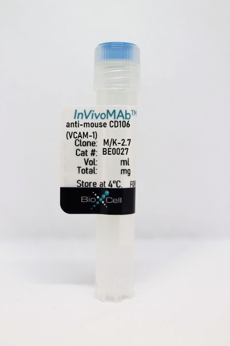
InVivoMAb anti-mouse CD106 (VCAM-1)
Product Details
The M/K-2.7 monoclonal antibody reacts with CD106 also known as VCAM-1 and INCAM-110. CD106 is a 110 kDa single chain type I glycoprotein that is expressed primarily on activated vascular endothelial cells but has also been reported on follicular and interfollicular dendritic cells, some macrophages, bone marrow stromal cells, and non-vascular cell populations within joints, kidney, muscle, heart, placenta, and brain. CD106 expression is induced by inflammatory stimuli and cytokines. CD106 binds the integrins CD49d/CD29 (VLA-4) and α4β7 which contribute to leukocyte adhesion, transmigration, and co-stimulation of T cell proliferation.Specifications
| Isotype | Rat IgG1, κ |
|---|---|
| Recommended Isotype Control(s) | InVivoMAb rat IgG1 isotype control, anti-horseradish peroxidase |
| Recommended Dilution Buffer | InVivoPure pH 7.0 Dilution Buffer |
| Conjugation | This product is unconjugated. Conjugation is available via our Antibody Conjugation Services. |
| Immunogen | Stromal cells from mouse bone marrow |
| Reported Applications |
in vivo VCAM-1 neutralization Immunofluorescence |
| Formulation |
PBS, pH 7.0 Contains no stabilizers or preservatives |
| Endotoxin |
<2EU/mg (<0.002EU/μg) Determined by LAL gel clotting assay |
| Purity |
>95% Determined by SDS-PAGE |
| Sterility | 0.2 µm filtration |
| Production | Purified from cell culture supernatant in an animal-free facility |
| Purification | Protein G |
| RRID | AB_1107572 |
| Molecular Weight | 150 kDa |
| Storage | The antibody solution should be stored at the stock concentration at 4°C. Do not freeze. |
Recommended Products
-
Recommended Isotype Control(s)
InVivoMAb rat IgG1 isotype control, anti-horseradish peroxidase
-

Recommended Dilution Buffer
InVivoPure pH 7.0 Dilution Buffer
in vivo VCAM-1 neutralization
Yousef, H., et al. (2019). "Aged blood impairs hippocampal neural precursor activity and activates microglia via brain endothelial cell VCAM1" Nat Med 25(6): 988-1000. PubMed
An aged circulatory environment can activate microglia, reduce neural precursor cell activity and impair cognition in mice. We hypothesized that brain endothelial cells (BECs) mediate at least some of these effects. We observe that BECs in the aged mouse hippocampus express an inflammatory transcriptional profile with focal upregulation of vascular cell adhesion molecule 1 (VCAM1), a protein that facilitates vascular-immune cell interactions. Concomitantly, levels of the shed, soluble form of VCAM1 are prominently increased in the plasma of aged humans and mice, and their plasma is sufficient to increase VCAM1 expression in cultured BECs and the hippocampi of young mice. Systemic administration of anti-VCAM1 antibody or genetic ablation of Vcam1 in BECs counteracts the detrimental effects of plasma from aged individuals on young brains and reverses aging aspects, including microglial reactivity and cognitive deficits, in the brains of aged mice. Together, these findings establish brain endothelial VCAM1 at the blood-brain barrier as a possible target to treat age-related neurodegeneration.
in vivo VCAM-1 neutralization
de Juan, A., et al. (2019). "Artery-Associated Sympathetic Innervation Drives Rhythmic Vascular Inflammation of Arteries and Veins" Circulation 140(13): 1100-1114. PubMed
BACKGROUND: The incidence of acute cardiovascular complications is highly time-of-day dependent. However, the mechanisms driving rhythmicity of ischemic vascular events are unknown. Although enhanced numbers of leukocytes have been linked to an increased risk of cardiovascular complications, the role that rhythmic leukocyte adhesion plays in different vascular beds has not been studied. METHODS: We evaluated leukocyte recruitment in vivo by using real-time multichannel fluorescence intravital microscopy of a tumor necrosis factor-α-induced acute inflammation model in both murine arterial and venous macrovasculature and microvasculature. These approaches were complemented with genetic, surgical, and pharmacological ablation of sympathetic nerves or adrenergic receptors to assess their relevance for rhythmic leukocyte adhesion. In addition, we genetically targeted the key circadian clock gene Bmal1 (also known as Arntl) in a lineage-specific manner to dissect the importance of oscillations in leukocytes and components of the vessel wall in this process. RESULTS: In vivo quantitative imaging analyses of acute inflammation revealed a 24-hour rhythm in leukocyte recruitment to arteries and veins of the mouse macrovasculature and microvasculature. Unexpectedly, although in arteries leukocyte adhesion was highest in the morning, it peaked at night in veins. This phase shift was governed by a rhythmic microenvironment and a vessel type-specific oscillatory pattern in the expression of promigratory molecules. Differences in cell adhesion molecules and leukocyte adhesion were ablated when disrupting sympathetic nerves, demonstrating their critical role in this process and the importance of β(2)-adrenergic receptor signaling. Loss of the core clock gene Bmal1 in leukocytes, endothelial cells, or arterial mural cells affected the oscillations in a vessel type-specific manner. Rhythmicity in the intravascular reactivity of adherent leukocytes resulted in increased interactions with platelets in the morning in arteries and in veins at night with a higher predisposition to acute thrombosis at different times as a consequence. CONCLUSIONS: Together, our findings point to an important and previously unrecognized role of artery-associated sympathetic innervation in governing rhythmicity in vascular inflammation in both arteries and veins and its potential implications in the occurrence of time-of-day-dependent vessel type-specific thrombotic events.
in vivo VCAM-1 neutralization
He, W., et al. (2018). "Circadian Expression of Migratory Factors Establishes Lineage-Specific Signatures that Guide the Homing of Leukocyte Subsets to Tissues" Immunity 49(6): 1175-1190.e1177. PubMed
The number of leukocytes present in circulation varies throughout the day, reflecting bone marrow output and emigration from blood into tissues. Using an organism-wide circadian screening approach, we detected oscillations in pro-migratory factors that were distinct for specific vascular beds and individual leukocyte subsets. This rhythmic molecular signature governed time-of-day-dependent homing behavior of leukocyte subsets to specific organs. Ablation of BMAL1, a transcription factor central to circadian clock function, in endothelial cells or leukocyte subsets demonstrated that rhythmic recruitment is dependent on both microenvironmental and cell-autonomous oscillations. These oscillatory patterns defined leukocyte trafficking in both homeostasis and inflammation and determined detectable tumor burden in blood cancer models. Rhythms in the expression of pro-migratory factors and migration capacities were preserved in human primary leukocytes. The definition of spatial and temporal expression profiles of pro-migratory factors guiding leukocyte migration patterns to organs provides a resource for the further study of the impact of circadian rhythms in immunity.
in vivo VCAM-1 neutralization
Kapitsinou, P. P., et al. (2014). "Endothelial HIF-2 mediates protection and recovery from ischemic kidney injury" J Clin Invest 124(6): 2396-2409. PubMed
The hypoxia-inducible transcription factors HIF-1 and HIF-2 mediate key cellular adaptions to hypoxia and contribute to renal homeostasis and pathophysiology; however, little is known about the cell type-specific functions of HIF-1 and HIF-2 in response to ischemic kidney injury. Here, we used a genetic approach to specifically dissect the roles of endothelial HIF-1 and HIF-2 in murine models of hypoxic kidney injury induced by ischemia reperfusion or ureteral obstruction. In both models, inactivation of endothelial HIF increased injury-associated renal inflammation and fibrosis. Specifically, inactivation of endothelial HIF-2alpha, but not endothelial HIF-1alpha, resulted in increased expression of renal injury markers and inflammatory cell infiltration in the postischemic kidney, which was reversed by blockade of vascular cell adhesion molecule-1 (VCAM1) and very late antigen-4 (VLA4) using monoclonal antibodies. In contrast, pharmacologic or genetic activation of HIF via HIF prolyl-hydroxylase inhibition protected wild-type animals from ischemic kidney injury and inflammation; however, these same protective effects were not observed in HIF prolyl-hydroxylase inhibitor-treated animals lacking endothelial HIF-2. Taken together, our data indicate that endothelial HIF-2 protects from hypoxia-induced renal damage and represents a potential therapeutic target for renoprotection and prevention of fibrosis following acute ischemic injury.
in vivo VCAM-1 neutralization
Chow, A., et al. (2013). "CD169(+) macrophages provide a niche promoting erythropoiesis under homeostasis and stress" Nat Med 19(4): 429-436. PubMed
A role for macrophages in erythropoiesis was suggested several decades ago when erythroblastic islands in the bone marrow, composed of a central macrophage surrounded by developing erythroblasts, were described. However, the in vivo role of macrophages in erythropoiesis under homeostatic conditions or in disease remains unclear. We found that specific depletion of CD169(+) macrophages markedly reduced the number of erythroblasts in the bone marrow but did not result in overt anemia under homeostatic conditions, probably because of concomitant alterations in red blood cell clearance. However, CD169(+) macrophage depletion significantly impaired erythropoietic recovery from hemolytic anemia, acute blood loss and myeloablation. Furthermore, macrophage depletion normalized the erythroid compartment in a JAK2(V617F)-driven mouse model of polycythemia vera, suggesting that erythropoiesis in polycythemia vera remains under the control of macrophages in the bone marrow and splenic microenvironments. These results indicate that CD169(+) macrophages promote late erythroid maturation and that modulation of the macrophage compartment may be a new strategy to treat erythropoietic disorders.
in vivo VCAM-1 neutralization, Immunofluorescence
Brinkman, C. C., et al. (2013). "Peripheral tissue homing receptors enable T cell entry into lymph nodes and affect the anatomical distribution of memory cells" J Immunol 191(5): 2412-2425. PubMed
Peripheral tissue homing receptors enable T cells to access inflamed nonlymphoid tissues. In this study, we show that two such molecules, E-selectin ligand and alpha4beta1 integrin, enable activated and memory T cells to enter lymph nodes (LN) as well. This affects the quantitative and qualitative distribution of these cells among regional LN beds. CD8 memory T cells in LN that express these molecules were mostly CD62L(lo) and would normally be classified as effector memory cells. However, similar to central memory cells, they expanded upon Ag re-encounter. This led to differences in the magnitude of the recall response that depended on the route of immunization. These novel cells share properties of both central and effector memory cells and reside in LN based on previously undescribed mechanisms of entry.
in vivo VCAM-1 neutralization
Thomas, S. Y., et al. (2011). "PLZF induces an intravascular surveillance program mediated by long-lived LFA-1-ICAM-1 interactions" J Exp Med 208(6): 1179-1188. PubMed
Innate-like NKT cells conspicuously accumulate within the liver microvasculature of healthy mice, crawling on the luminal side of endothelial cells, but their general recirculation pattern and the mechanism of their intravascular behavior have not been elucidated. Using parabiotic mice, we demonstrated that, despite their intravascular location, most liver NKT cells failed to recirculate. Antibody blocking experiments established that they were retained locally through constitutive LFA-1-intercellular adhesion molecule (ICAM) 1 interactions. This unprecedented lifelong intravascular residence could be induced in conventional CD4 T cells by the sole expression of promyelocytic leukemia zinc finger (PLZF), a transcription factor specifically expressed in the NKT lineage. These findings reveal the unique genetic and biochemical pathway that underlies the innate intravascular surveillance program of NKT cells.
- Immunology and Microbiology,
Vascular Cell Adhesion Molecule-1 (VCAM-1) contributes to macular fibrosis in neovascular age-related macular degeneration through modulating macrophage functions.
In Immunity & Ageing : I & A on 20 November 2023 by Deng, W., Yi, C., et al.
PubMed
Neovascular age-related macular degeneration (nAMD) is a major cause of blindness in the elderly. The disease is due to the growth of abnormal blood vessels into the macula, leading to the loss of central vision. Intravitreal injection of vascular endothelial growth factor (VEGF) inhibitors (e.g., anti-VEGF) is the standard of care for nAMD. However, nearly 50% of patients do not respond or respond poorly to the therapy. More importantly, up to 70% of nAMD patients develop macular fibrosis after 10 years of anti-VEGF therapy. The underlying mechanism of nAMD-mediated macular fibrosis is unknown although inflammation is known to play an important role in the development of abnormal macular blood vessels and its progression to fibro-vascular membrane. In this study, we measured the intraocular levels of adhesion molecule VCAM-1, ICAM-1, CD44, CD62L, and CD62P in nAMD patients with and without macular fibrosis and investigated the link between the levels of adhesion molecule and clinical features (e.g., visual improvement, retinal thickness, etc.). We further investigated the effect of VCAM-1 in macrophage function in vitro and the development of subretinal fibrosis in vivo using a two-stage laser-induced protocol. The aqueous levels of ICAM-1, VCAM-1, CD44, and CD62L were significantly higher in nAMD patients compared to cataract controls. The aqueous level of VCAM-1 (but not other adhesion molecules) was significantly higher in patients with macular fibrosis than those without and the level correlated positively with the retinal thickness. VCAM-1 was highly expressed at the lesion site in the mouse model of subretinal fibrosis. Blocking VCAM-1 or its receptor VLA-4 significantly prevented macrophage infiltration and reduced subretinal fibrosis in vivo. VCAM-1 induced macrophage migration and upregulated the expression of Arg-1, Mmp12 and Il6 but down-regulated the expression of iNOS and Il1b in macrophages. VCAM-1 may contribute to the development of macular fibrosis in nAMD patients by modulating macrophage functions, including migration and profibrotic polarization. © 2023. The Author(s).
- Cancer Research,
- Immunology and Microbiology,
- Stem Cells and Developmental Biology,
- In Vivo,
- Mus musculus (House mouse)
Pericyte stem cells induce Ly6G+ cell accumulation and immunotherapy resistance in pancreatic cancer.
In EMBO Reports on 5 April 2023 by Wu, Z., Thierry, K., et al.
PubMed
We report the identification of a cell population that shares pericyte, stromal and stemness features, does not harbor the KrasG12D mutation and drives tumoral growth in vitro and in vivo. We term these cells pericyte stem cells (PeSCs) and define them as CD45- EPCAM- CD29+ CD106+ CD24+ CD44+ cells. We perform studies with p48-Cre;KrasG12D (KC), pdx1-Cre;KrasG12D ;Ink4a/Arffl/fl (KIC) and pdx1-Cre;KrasG12D ;p53R172H (KPC) and tumor tissues from PDAC and chronic pancreatitis patients. We also perform single-cell RNAseq analysis and reveal a unique signature of PeSC. Under steady-state conditions, PeSCs are barely detectable in the pancreas but present in the neoplastic microenvironment both in humans and mice. The coinjection of PeSCs and tumor epithelial cells leads to increased tumor growth, differentiation of Ly6G+ myeloid-derived suppressor cells, and a decreased amount of F4/80+ macrophages and CD11c+ dendritic cells. This population induces resistance to anti-PD-1 immunotherapy when coinjected with epithelial tumor cells. Our data reveal the existence of a cell population that instructs immunosuppressive myeloid cell responses to bypass PD-1 targeting and thus suggest potential new approaches for overcoming resistance to immunotherapy in clinical settings. © 2023 The Authors. Published under the terms of the CC BY NC ND 4.0 license.
- Cancer Research,
- Mus musculus (House mouse)
Spatiotemporal regulation of cholangiocarcinoma growth and dissemination by peritumoral myofibroblasts in a Vcam1-dependent manner.
In Oncogene on 1 April 2023 by Tian, C., Li, L., et al.
PubMed
Intrahepatic cholangiocarcinoma (iCCA) is characterized by its highly desmoplastic stroma. Myofibroblasts (MFs) are present both within the tumor mass (intratumoral MFs, iMFs) and at the tumor border (peritumoral MFs, pMFs). Using a spheroid-based coculture system, we show that the initial iCCA-pMF contact is growth suppressive to the tumor cells. However, prolonged iCCA-pMF interaction elicits significant tumor cell invasion and dissemination. We find that vascular cell adhesion molecule-1 (Vcam1) level is elevated in tumor cells in contact with pMFs but low in disseminated tumor cells both in vitro and in vivo. A gene regulatory network analysis of mouse and patient iCCA tumors and Vcam1 knockout (Vcam1KO) demonstrate a heavy involvement of Vcam1 in epithelial-to-mesenchymal transition. While Vcam1KO has only a limited impact on tumor cell growth in their monoculture, Vcam1KO spheroids exhibit instant dissemination and a severe growth defect when cocultured with pMFs. When transplanted into the liver, Vcam1KO iCCA cells show a similar increase in dissemination but a significant defect in establishing primary and metastatic tumors. Incomplete blocking of Vcam1 in vivo reduces the size but increase the number of metastatic lesions. Overall, our study shows a spatiotemporal regulation of iCCA growth and dissemination by pMFs in a Vcam1-dependent manner. © 2023. The Author(s).
- Cancer Research
Spatiotemporal Dynamics of Vcam1 Regulates Cholangiocarcinoma Mass Expansion and Tumor Dissemination under Growth-suppressive Peritumoral Myofibroblasts
Preprint on BioRxiv : the Preprint Server for Biology on 24 January 2023 by Tian, C., Li, L., et al.
PubMed
ABSTRACT Intrahepatic cholangiocarcinoma (iCCA) is characterized by its highly desmoplastic stroma. Myofibroblasts (MFs) are present both within the tumor mass (intratumoral MFs, iMFs) and at the tumor border (peritumoral MFs, pMFs). Using a spheroid-based coculture system, we show that the initial iCCA-pMF contact is growth suppressive to the tumor cells. However, prolonged iCCA-pMF interaction elicits significant tumor cell invasion and dissemination. We find that vascular cell adhesion molecule-1 ( Vcam1 ) level is elevated in tumor cells in contact with pMFs but low in disseminated tumor cells both in vitro and in vivo. A gene regulatory network analysis of mouse and patient iCCA tumors and Vcam1 knockout ( Vcam1 KO ) demonstrate a heavy involvement of Vcam1 in epithelial-to-mesenchymal transition. While Vcam1 KO has only a limited impact on tumor cell growth in their monoculture, Vcam1 KO spheroids exhibit instant dissemination and a severe growth defect when cocultured with pMFs. When transplanted into the liver, Vcam1 KO iCCA cells show a similar increase in dissemination but a significant defect in establishing primary and metastatic tumors. Incomplete blocking of Vcam1 in vivo reduces the size but increase the number of metastatic lesions. Overall, our study shows a spatiotemporal regulation of iCCA growth and dissemination by pMFs in a Vcam1-dependent manner.
- In Vivo,
- Mus musculus (House mouse),
- Cardiovascular biology,
- Immunology and Microbiology,
- Pharmacology
Blocking VCAM-1 ameliorates hypertensive cardiac remodeling by impeding macrophage infiltration.
In Frontiers in Pharmacology on 6 December 2022 by Qiu, Z. Y., Yu, W. J., et al.
PubMed
Cardiac remodeling is an important mechanism of heart failure, which frequently results from leukocyte infiltration. Vascular cellular adhesion molecule-1 (VCAM-1) plays a critical role in leukocyte adhesion and transmigration. However, the importance of VCAM-1 in the development of angiotensin II (Ang II)-induced cardiac remodeling remains unclear. Wild-type (WT) mice were infused with Ang II (1,000 ng/kg/min) for 14 days and simultaneously treated with VCAM-1 neutralizing antibody (0.1 or 0.2 mg) or IgG control. Systolic blood pressure (SBP) and cardiac function were detected by a tail-cuff and echocardiography. Cardiac remodeling was evaluated by histological staining. Adhesion and migration of bone marrow macrophages (BMMs) were evaluated in vitro. Our results indicated that VCAM-1 levels were increased in the serum of patients with heart failure (HF) and the hearts of Ang II-infused mice. Furthermore, Ang II-caused hypertension, cardiac dysfunction, hypertrophy, fibrosis, infiltration of VLA-4+ BMMs and oxidative stress were dose-dependently attenuated in mice administered VCAM-1 neutralizing antibody. In addition, blocking VCAM-1 markedly alleviated Ang II-induced BMMs adhesion and migration, therefore inhibited cardiomyocyte hypertrophy and fibroblast activation. In conclusion, the data reveal that blocking VCAM-1 ameliorates hypertensive cardiac remodeling by impeding VLA-4+ macrophage infiltration. Selective blockage of VCAM-1 may be a novel therapeutic strategy for hypertensive cardiac diseases. Copyright © 2022 Qiu, Yu, Bai and Lin.
- In Vivo,
- Mus musculus (House mouse),
- Cardiovascular biology,
- Pharmacology
Blocking VCAM-1 Prevents Angiotensin II-Induced Hypertension and Vascular Remodeling in Mice.
In Frontiers in Pharmacology on 1 March 2022 by Yin, L., Bai, J., et al.
PubMed
Adhesion of monocytes to the vascular endothelium frequently leads to an inflammatory response, which contributes to hypertension and vascular remodeling. Vascular cellular adhesion molecule-1 (VCAM-1) plays an important role in leukocyte adhesion and migration during inflammatory diseases. However, its role in angiotensin (Ang) II -induced hypertension and vascular dysfunction remains largely unknown. Wild-type (WT) mice were administered a VCAM-1 neutralizing antibody (0.1 or 0.2 mg/mouse/day) or IgG control and then infused with Ang II (490 ng kg-1 min-1) or saline continuously for 14 days. Systolic blood pressure (SBP) was measured with a tail-cuff system, pathological changes in the aorta were assessed by histological staining, and vascular relaxation was analyzed an aortic ring assay. Our results indicated that compared with saline infusion, Ang II infusion significantly upregulated VCAM-1 expression in the mouse aorta and serum. Moreover, Ang II infusion markedly increased arterial hypertension, wall thickness, fibrosis, infiltration of Mac-2+ macrophages, reactive oxygen species (ROS) production and vascular relaxation dysfunction. Conversely, blockade of VCAM-1 with a neutralizing antibody substantially alleviated these effects. In vitro experiments further confirmed that the VCAM-1 neutralizing antibody inhibited Ang II-induced macrophage adhesion and migration and DNA damage and oxidative stress in endothelial cells (ECs). In conclusion, these results indicate that blockade of VCAM-1 exerts a protective effect against Ang II-induced arterial hypertension and dysfunction by regulating monocytes adhesion and infiltration into the endothelium and represents a novel therapeutic approach for hypertension. Copyright © 2022 Yin, Bai, Yu, Liu, Li and Lin.
- In Vivo,
- Mus musculus (House mouse)
Brain Endothelial Cells Are Exquisite Sensors of Age-Related Circulatory Cues.
In Cell Reports on 31 March 2020 by Chen, M. B., Yang, A. C., et al.
PubMed
Brain endothelial cells (BECs) are key constituents of the blood-brain barrier (BBB), protecting the brain from pathogens and restricting access of circulatory factors. Yet, because circulatory proteins have prominent age-related effects on adult neurogenesis, neuroinflammation, and cognitive function in mice, we wondered whether BECs receive and potentially relay signals between the blood and brain. Using single-cell RNA sequencing of hippocampal BECs, we discover that capillary BECs-compared with arterial and venous BECs-undergo the greatest transcriptional changes in normal aging, upregulating innate immunity and oxidative stress response pathways. Short-term infusions of aged plasma into young mice recapitulate key aspects of this aging transcriptome, and remarkably, infusions of young plasma into aged mice exert rejuvenation effects on the capillary transcriptome. Together, these findings suggest that the transcriptional age of BECs is exquisitely sensitive to age-related circulatory cues and pinpoint the BBB itself as a promising therapeutic target to treat brain disease. Published by Elsevier Inc.
- In Vivo,
- Block,
- Mus musculus (House mouse),
- Cardiovascular biology,
- Immunology and Microbiology
Artery-Associated Sympathetic Innervation Drives Rhythmic Vascular Inflammation of Arteries and Veins.
In Circulation on 24 September 2019 by De Juan, A., Ince, L. M., et al.
PubMed
The incidence of acute cardiovascular complications is highly time-of-day dependent. However, the mechanisms driving rhythmicity of ischemic vascular events are unknown. Although enhanced numbers of leukocytes have been linked to an increased risk of cardiovascular complications, the role that rhythmic leukocyte adhesion plays in different vascular beds has not been studied.We evaluated leukocyte recruitment in vivo by using real-time multichannel fluorescence intravital microscopy of a tumor necrosis factor-α-induced acute inflammation model in both murine arterial and venous macrovasculature and microvasculature. These approaches were complemented with genetic, surgical, and pharmacological ablation of sympathetic nerves or adrenergic receptors to assess their relevance for rhythmic leukocyte adhesion. In addition, we genetically targeted the key circadian clock gene Bmal1 (also known as Arntl) in a lineage-specific manner to dissect the importance of oscillations in leukocytes and components of the vessel wall in this process.In vivo quantitative imaging analyses of acute inflammation revealed a 24-hour rhythm in leukocyte recruitment to arteries and veins of the mouse macrovasculature and microvasculature. Unexpectedly, although in arteries leukocyte adhesion was highest in the morning, it peaked at night in veins. This phase shift was governed by a rhythmic microenvironment and a vessel type-specific oscillatory pattern in the expression of promigratory molecules. Differences in cell adhesion molecules and leukocyte adhesion were ablated when disrupting sympathetic nerves, demonstrating their critical role in this process and the importance of β2-adrenergic receptor signaling. Loss of the core clock gene Bmal1 in leukocytes, endothelial cells, or arterial mural cells affected the oscillations in a vessel type-specific manner. Rhythmicity in the intravascular reactivity of adherent leukocytes resulted in increased interactions with platelets in the morning in arteries and in veins at night with a higher predisposition to acute thrombosis at different times as a consequence.Together, our findings point to an important and previously unrecognized role of artery-associated sympathetic innervation in governing rhythmicity in vascular inflammation in both arteries and veins and its potential implications in the occurrence of time-of-day-dependent vessel type-specific thrombotic events.
- In Vivo,
- FC/FACS,
- Mus musculus (House mouse),
- Cardiovascular biology,
- Neuroscience
Aged blood impairs hippocampal neural precursor activity and activates microglia via brain endothelial cell VCAM1.
In Nature Medicine on 1 June 2019 by Yousef, H., Czupalla, C. J., et al.
PubMed
An aged circulatory environment can activate microglia, reduce neural precursor cell activity and impair cognition in mice. We hypothesized that brain endothelial cells (BECs) mediate at least some of these effects. We observe that BECs in the aged mouse hippocampus express an inflammatory transcriptional profile with focal upregulation of vascular cell adhesion molecule 1 (VCAM1), a protein that facilitates vascular-immune cell interactions. Concomitantly, levels of the shed, soluble form of VCAM1 are prominently increased in the plasma of aged humans and mice, and their plasma is sufficient to increase VCAM1 expression in cultured BECs and the hippocampi of young mice. Systemic administration of anti-VCAM1 antibody or genetic ablation of Vcam1 in BECs counteracts the detrimental effects of plasma from aged individuals on young brains and reverses aging aspects, including microglial reactivity and cognitive deficits, in the brains of aged mice. Together, these findings establish brain endothelial VCAM1 at the blood-brain barrier as a possible target to treat age-related neurodegeneration.
- In Vivo,
- Mus musculus (House mouse),
- Immunology and Microbiology
Interferon-γ facilitated adjuvant-induced arthritis at early stage.
In Scandinavian Journal of Immunology on 1 May 2019 by Ma, H., Xu, M., et al.
PubMed
Interferon-γ (IFN-γ) is a versatile cytokine which broadly involves in the inflammatory diseases, mediating both immune activation and tolerance. Here, we aimed to investigate the role of IFN-γ in the initiation of adjuvant-induced arthritis (AIA). In an AIA mice model, increasing IFN-γ mRNA was observed at day 3 and peaked on day 7. At day 3, the majority of IFN-γ-producing cells were located around vessels observed by immunofluorescent staining. Recombinant IFN-γ or anti-IFN-γ antibody was injected into the AIA paw on day 2 to study the outcome of AIA. The recipients of IFN-γ showed increased synovial inflammation, whereas anti-IFN-γ antibody injection repressed the expansion of inflammatory cells. As the percentages of blood monocytes were approximately equivalent, we hypothesized that IFN-γ might impact the access of innate leucocytes from blood to expand local inflammation at this stage. Analysis of tissue CD31 and vascular cell adhesion molecule-1 (VCAM-1) expressions suggested a positive effect of these factors in the development of inflammation, and IFN-γ affected the VCAM-1 expression. To further verify this idea, mice regionally injected with IFN-γ were systematically administrated with anti-VCAM-1 antibody during AIA induction. The IFN-γ expression was inhibited, and the development of AIA was partly abolished in these mice regardless of regional IFN-γ injection. These data suggested that IFN-γ might be critical for the expansion of AIA at early stage through helping inflammatory cell access. © 2019 The Foundation for the Scandinavian Journal of Immunology.
- In Vivo,
- Block,
- Mus musculus (House mouse),
- Immunology and Microbiology
Circadian Expression of Migratory Factors Establishes Lineage-Specific Signatures that Guide the Homing of Leukocyte Subsets to Tissues.
In Immunity on 18 December 2018 by He, W., Holtkamp, S., et al.
PubMed
The number of leukocytes present in circulation varies throughout the day, reflecting bone marrow output and emigration from blood into tissues. Using an organism-wide circadian screening approach, we detected oscillations in pro-migratory factors that were distinct for specific vascular beds and individual leukocyte subsets. This rhythmic molecular signature governed time-of-day-dependent homing behavior of leukocyte subsets to specific organs. Ablation of BMAL1, a transcription factor central to circadian clock function, in endothelial cells or leukocyte subsets demonstrated that rhythmic recruitment is dependent on both microenvironmental and cell-autonomous oscillations. These oscillatory patterns defined leukocyte trafficking in both homeostasis and inflammation and determined detectable tumor burden in blood cancer models. Rhythms in the expression of pro-migratory factors and migration capacities were preserved in human primary leukocytes. The definition of spatial and temporal expression profiles of pro-migratory factors guiding leukocyte migration patterns to organs provides a resource for the further study of the impact of circadian rhythms in immunity.Copyright © 2018 The Author(s). Published by Elsevier Inc. All rights reserved.
- In Vivo,
- Mus musculus (House mouse),
- Cancer Research,
- Cardiovascular biology
Kindlin-1 Promotes Pulmonary Breast Cancer Metastasis.
In Cancer Research on 15 March 2018 by Sarvi, S., Patel, H., et al.
PubMed
In breast cancer, increased expression of the cytoskeletal adaptor protein Kindlin-1 has been linked to increased risks of lung metastasis, but the functional basis is unknown. Here, we show that in a mouse model of polyomavirus middle T antigen-induced mammary tumorigenesis, loss of Kindlin-1 reduced early pulmonary arrest and later development of lung metastasis. This phenotype relied on the ability of Kindlin-1 to bind and activate β integrin heterodimers. Kindlin-1 loss reduced α4 integrin-mediated adhesion of mammary tumor cells to the adhesion molecule VCAM-1 on endothelial cells. Treating mice with an anti-VCAM-1 blocking antibody prevented early pulmonary arrest. Kindlin-1 loss also resulted in reduced secretion of several factors linked to metastatic spread, including the lung metastasis regulator tenascin-C, showing that Kindlin-1 regulated metastatic dissemination by an additional mechanism in the tumor microenvironment. Overall, our results show that Kindlin-1 contributes functionally to early pulmonary metastasis of breast cancer.Significance: These findings provide a mechanistic proof in mice that Kindin-1, an integrin-binding adaptor protein, is a critical mediator of early lung metastasis of breast cancer. Cancer Res; 78(6); 1484-96. ©2018 AACR. ©2018 American Association for Cancer Research.
- FC/FACS,
- Cardiovascular biology,
- Neuroscience,
- Mus musculus (House mouse)
Aged blood inhibits hippocampal neurogenesis and activates microglia through VCAM1 at the blood-brain barrier
Preprint on BioRxiv : the Preprint Server for Biology on 3 January 2018 by Yousef, H., Czupalla, C. J., et al.
PubMed
An aged circulatory environment can promote brain dysfunction and we hypothesized that the blood-brain barrier (BBB) mediates at least some of these effects. We observe brain endothelial cells (BECs) in the aged mouse hippocampus express an inflammatory transcriptional profile with focal upregulation of Vascular Cell Adhesion Molecule 1 (VCAM1), a protein that facilitates vascular-immune cell interactions. Concomitantly, the shed, soluble form of VCAM1 is prominently increased in the aged circulation of humans and mice, and aged plasma is sufficient to increase VCAM1 expression in cultured BECs and young mouse hippocampi. Systemic anti-VCAM1 antibody or genetic ablation of VCAM1 in BECs counteracts the detrimental effects of aged plasma on young brains and reverses aging aspects in old mouse brains. Thus, VCAM1 is a negative regulator of adult neurogenesis and inducer of microglial reactivity, establishing VCAM1 and the luminal side of the BBB as possible targets to treat age-related neurodegeneration.
- In Vivo,
- Mus musculus (House mouse)
The retinal pigment epithelium as a gateway for monocyte trafficking into the eye.
In The EMBO Journal on 1 June 2016 by Benhar, I., Reemst, K., et al.
PubMed
The choroid plexus epithelium within the brain ventricles orchestrates blood-derived monocyte entry to the central nervous system under injurious conditions, including when the primary injury site is remote from the brain. Here, we hypothesized that the retinal pigment epithelium (RPE) serves a parallel role, as a gateway for monocyte trafficking to the retina following direct or remote injury. We found elevated expression of genes encoding leukocyte trafficking determinants in mouse RPE as a consequence of retinal glutamate intoxication or optic nerve crush (ONC). Blocking VCAM-1 after ONC interfered with monocyte infiltration into the retina and resulted in a local pro-inflammatory cytokine bias. Live imaging of the injured eye showed monocyte accumulation first in the RPE, and subsequently in the retina, and peripheral leukocytes formed close contact with the RPE Our findings further implied that the ocular milieu can confer monocytes a phenotype advantageous for neuroprotection. These results suggest that the eye utilizes a mechanism of crosstalk with the immune system similar to that of the brain, whereby epithelial barriers serve as gateways for leukocyte entry. © 2016 The Authors.
- In Vivo,
- Mus musculus (House mouse)
Endothelial HIF-2 mediates protection and recovery from ischemic kidney injury.
In The Journal of Clinical Investigation on 1 June 2014 by Kapitsinou, P. P., Sano, H., et al.
PubMed
The hypoxia-inducible transcription factors HIF-1 and HIF-2 mediate key cellular adaptions to hypoxia and contribute to renal homeostasis and pathophysiology; however, little is known about the cell type-specific functions of HIF-1 and HIF-2 in response to ischemic kidney injury. Here, we used a genetic approach to specifically dissect the roles of endothelial HIF-1 and HIF-2 in murine models of hypoxic kidney injury induced by ischemia reperfusion or ureteral obstruction. In both models, inactivation of endothelial HIF increased injury-associated renal inflammation and fibrosis. Specifically, inactivation of endothelial HIF-2α, but not endothelial HIF-1α, resulted in increased expression of renal injury markers and inflammatory cell infiltration in the postischemic kidney, which was reversed by blockade of vascular cell adhesion molecule-1 (VCAM1) and very late antigen-4 (VLA4) using monoclonal antibodies. In contrast, pharmacologic or genetic activation of HIF via HIF prolyl-hydroxylase inhibition protected wild-type animals from ischemic kidney injury and inflammation; however, these same protective effects were not observed in HIF prolyl-hydroxylase inhibitor-treated animals lacking endothelial HIF-2. Taken together, our data indicate that endothelial HIF-2 protects from hypoxia-induced renal damage and represents a potential therapeutic target for renoprotection and prevention of fibrosis following acute ischemic injury.
- Block,
- Mus musculus (House mouse)
CD169⁺ macrophages provide a niche promoting erythropoiesis under homeostasis and stress.
In Nature Medicine on 1 April 2013 by Chow, A., Huggins, M., et al.
PubMed
A role for macrophages in erythropoiesis was suggested several decades ago when erythroblastic islands in the bone marrow, composed of a central macrophage surrounded by developing erythroblasts, were described. However, the in vivo role of macrophages in erythropoiesis under homeostatic conditions or in disease remains unclear. We found that specific depletion of CD169(+) macrophages markedly reduced the number of erythroblasts in the bone marrow but did not result in overt anemia under homeostatic conditions, probably because of concomitant alterations in red blood cell clearance. However, CD169(+) macrophage depletion significantly impaired erythropoietic recovery from hemolytic anemia, acute blood loss and myeloablation. Furthermore, macrophage depletion normalized the erythroid compartment in a JAK2(V617F)-driven mouse model of polycythemia vera, suggesting that erythropoiesis in polycythemia vera remains under the control of macrophages in the bone marrow and splenic microenvironments. These results indicate that CD169(+) macrophages promote late erythroid maturation and that modulation of the macrophage compartment may be a new strategy to treat erythropoietic disorders.