Catalog #CP023
RecombiMAb anti-mouse PD-L2 (B7-DC)
Clone
TY25-CP023
Reactivities
Mouse
Isotype
Mouse IgG2a, κ
(switched from Rat Ig2a, κ)
(switched from Rat Ig2a, κ)
You may also be interested in:
Product Description
The TY25-CP023 monoclonal antibody is a recombinant, Fc-engineered chimeric version of the original TY25 antibody. The variable domain sequences are identical but the constant region sequences have been switched from Rat IgG2a, κ to Mouse IgG2a, κ for use in murine models. Species-matched chimeric antibodies exhibit regulated effector functions—including Fc receptor binding and complement activation—and result in less immunogenicity and formation of anti-drug antibodies (ADAs) than xenogenic antibodies in animal models. This antibody has an effector function competent Fc domain allowing for activation of Fcγ receptors (FcγRs) to trigger antibody‑dependent cellular cytotoxicity (ADCC), antibody‑dependent cellular phagocytosis (ADCP), complement‑dependent cytotoxicity (CDC) and opsonization to promote target cell depletion. The mouse IgG2a isotype demonstrates strong effector functions due to potent interaction with mFcγRIV, which is functionally similar to the FcγRIIIa receptor involved in human ADCC. The highly controlled sequence and lack of genetic drift in recombinant antibodies provide more reliable and reproducible results over hybridoma derived antibodies.
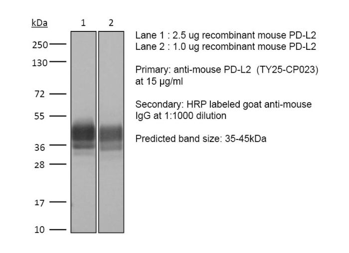
The TY25-CP023 monoclonal antibody reacts with mouse PD-L2 (programmed death ligand 2) also known as B7-DC or CD273. PD-L2 is a 25 kDa type I transmembrane protein that belongs to the B7 family of the Ig superfamily. PD-L2 is expressed on monocytes, macrophages and subsets of dendritic cells. PD-L2 binds to its receptor, PD-1, found on CD4 and CD8 thymocytes as well as activated T and B lymphocytes and myeloid cells. Engagement of PD-L2 with PD-1 leads to inhibition of TCR-mediated T cell proliferation and cytokine production. The TY25 antibody has been reported to block PD-1 mediated interactions in vivo.
The TY25-CP023 monoclonal antibody reacts with mouse PD-L2 (programmed death ligand 2) also known as B7-DC or CD273. PD-L2 is a 25 kDa type I transmembrane protein that belongs to the B7 family of the Ig superfamily. PD-L2 is expressed on monocytes, macrophages and subsets of dendritic cells. PD-L2 binds to its receptor, PD-1, found on CD4 and CD8 thymocytes as well as activated T and B lymphocytes and myeloid cells. Engagement of PD-L2 with PD-1 leads to inhibition of TCR-mediated T cell proliferation and cytokine production. The TY25 antibody has been reported to block PD-1 mediated interactions in vivo.
Specifications
| Isotype | Mouse IgG2a, κ |
|---|---|
| Recommended Isotype Control(s) | RecombiMAb mouse IgG2a isotype control, anti-hen egg lysozyme |
| Recommended Dilution Buffer | InVivoPure pH 7.0 Dilution Buffer |
| Immunogen | Mouse PD-L2 transfected cell line |
| Reported Applications |
in vivo PD-L2 blockade* in vitro PD-L2 blockade* Immunohistochemistry (frozen)* Flow cytometry* *Reported for the original Rat IgG2a, κ antibody. For information on in vivo applications, please contact technicalservice@bioxcell.com |
| Formulation |
PBS, pH 7.0 Contains no stabilizers or preservatives |
| Endotoxin |
≤0.5EU/mg (≤0.0005EU/μg) Determined by LAL assay |
| Aggregation |
<5% Determined by SEC |
| Purity |
≥95% Determined by SDS-PAGE |
| Sterility | 0.2 µm filtration |
| Production | Purified from mammalian cell supernatant in an animal-free facility |
| Purification | Protein G |
| Molecular Weight | 150 kDa |
| Murine Pathogen Tests |
Ectromelia/Mousepox Virus: Negative Hantavirus: Negative K Virus: Negative Lactate Dehydrogenase-Elevating Virus: Negative Lymphocytic Choriomeningitis virus: Negative Mouse Adenovirus: Negative Mouse Cytomegalovirus: Negative Mouse Hepatitis Virus: Negative Mouse Minute Virus: Negative Mouse Norovirus: Negative Mouse Parvovirus: Negative Mouse Rotavirus: Negative Mycoplasma Pulmonis: Negative Pneumonia Virus of Mice: Negative Polyoma Virus: Negative Reovirus Screen: Negative Sendai Virus: Negative Theiler’s Murine Encephalomyelitis: Negative |
| Storage | The antibody solution should be stored at the stock concentration at 4°C. Do not freeze. |
| Need a Custom Formulation? | See All Antibody Customization Options |