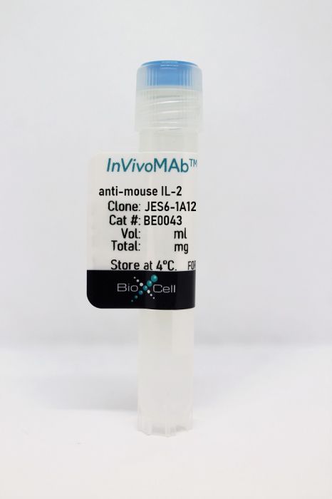
InVivoMAb anti-mouse IL-2
Product Details
The JES6-1A12 monoclonal antibody reacts with mouse IL-2 a 17 kDa cytokine that is mainly produced by T cells in response to antigenic or mitogenic stimulation. IL-2 is required for T cell proliferation and other activities crucial to the regulation of immunity. The cytokine can also stimulate the growth and differentiation of B cells monocytes/macrophages and NK cells. Additionally IL-2 prevents autoimmune diseases by promoting the differentiation of certain immature T cells into regulatory T cells. The JES6-1A12 antibody has been shown to neutralize IL-2 in vivo .Specifications
| Isotype | Rat IgG2a, κ |
|---|---|
| Recommended Isotype Control(s) | InVivoMAb rat IgG2a isotype control, anti-trinitrophenol |
| Recommended Dilution Buffer | InVivoPure pH 7.0 Dilution Buffer |
| Conjugation | This product is unconjugated. Conjugation is available via our Antibody Conjugation Services. |
| Immunogen | Recombinant mouse IL-2 |
| Reported Applications |
in vivo IL-2 neutralization in vivo IL-2 receptor stimulation (as a complex with IL-2) |
| Formulation |
PBS, pH 7.0 Contains no stabilizers or preservatives |
| Endotoxin |
<2EU/mg (<0.002EU/μg) Determined by LAL gel clotting assay |
| Purity |
>95% Determined by SDS-PAGE |
| Sterility | 0.2 μM filtered |
| Production | Purified from cell culture supernatant in an animal-free facility |
| Purification | Protein G |
| RRID | AB_1107702 |
| Molecular Weight | 150 kDa |
| Storage | The antibody solution should be stored at the stock concentration at 4°C. Do not freeze. |
Recommended Products
in vivo IL-2 receptor stimulation (as a complex with IL-2)
Littwitz-Salomon, E., et al. (2015). "Activated regulatory T cells suppress effector NK cell responses by an IL-2-mediated mechanism during an acute retroviral infection" Retrovirology 12: 66. PubMed
BACKGROUND: It is well established that effector T cell responses are crucial for the control of most virus infections, but they are often tightly controlled by regulatory T cells (Treg) to minimize immunopathology. NK cells also contribute to virus control but it is not known if their antiviral effect is influenced by virus-induced Tregs as well. We therefore analyzed whether antiretroviral NK cell functions are inhibited by Tregs during an acute Friend retrovirus infection of mice. RESULTS: Selective depletion of Tregs by using the transgenic DEREG mouse model resulted in improved NK cell proliferation, maturation and effector cell differentiation. Suppression of NK cell functions depended on IL-2 consumption by Tregs, which could be overcome by specific NK cell stimulation with an IL-2/anti-IL-2 mAb complex. CONCLUSIONS: The current study demonstrates that virus-induced Tregs indeed inhibit antiviral NK cell responses and describes a targeted immunotherapy that can abrogate the suppression of NK cells by Tregs.
in vivo IL-2 receptor stimulation (as a complex with IL-2)
Bouchery, T., et al. (2015). "ILC2s and T cells cooperate to ensure maintenance of M2 macrophages for lung immunity against hookworms" Nat Commun 6: 6970. PubMed
Defining the immune mechanisms underlying protective immunity to helminth infection remains an important challenge. Here we report that lung CD4(+) T cells and Group 2 innate lymphoid cells (ILC2s) work in concert to block Nippostrongylus brasiliensis (Nb) development in the parenchyma within 48 h in mice. Immune-damaged larvae have a striking morphological defect that is dependent on the expansion of IL-13-producing ILC2 and CD4(+) T cells, and the activation of M2 macrophages. This T-cell requirement can be bypassed by administration of IL-2 or IL-33, resulting in expansion of IL-13-producing ILC2s and larval killing. Depletion of ILC2s inhibits larval killing in IL-2-treated mice. Our results broaden understanding of ILC2’s role in immunity to helminths by demonstrating that they not only act as alarmin sensors, but can also be sustained by CD4(+) T cells, ensuring both the prompt activation and the maintenance of IL-13-dependent M2 macrophage immunity in the lung.
in vivo IL-2 neutralization
Joedicke, J. J., et al. (2014). "Activated CD8+ T cells induce expansion of Vbeta5+ regulatory T cells via TNFR2 signaling" J Immunol 193(6): 2952-2960. PubMed
Vbeta5(+) regulatory T cells (Tregs), which are specific for a mouse endogenous retroviral superantigen, become activated and proliferate in response to Friend virus (FV) infection. We previously reported that FV-induced expansion of this Treg subset was dependent on CD8(+) T cells and TNF-alpha, but independent of IL-2. We now show that the inflammatory milieu associated with FV infection is not necessary for induction of Vbeta5(+) Treg expansion. Rather, it is the presence of activated CD8(+) T cells that is critical for their expansion. The data indicate that the mechanism involves signaling between the membrane-bound form of TNF-alpha on activated CD8(+) T cells and TNFR2 on Tregs. CD8(+) T cells expressing membrane-bound TNF-alpha but no soluble TNF-alpha remained competent to induce strong Vbeta5(+) Treg expansion in vivo. In addition, Vbeta5(+) Tregs expressing only TNFR2 but no TNFR1 were still responsive to expansion. Finally, treatment of naive mice with soluble TNF-alpha did not induce Vbeta5(+) Treg expansion, but treatment with a TNFR2-specific agonist did. These results reveal a new mechanism of intercellular communication between activated CD8(+) T cell effectors and Tregs that results in the activation and expansion of a Treg subset that subsequently suppresses CD8(+) T cell functions.
in vivo IL-2 neutralization
Marshall, D., et al. (2014). "Differential requirement for IL-2 and IL-15 during bifurcated development of thymic regulatory T cells" J Immunol 193(11): 5525-5533. PubMed
The developmental pathways of regulatory T cells (T(reg)) generation in the thymus are not fully understood. In this study, we reconstituted thymic development of Zap70-deficient thymocytes with a tetracycline-inducible Zap70 transgene to allow temporal dissection of T(reg) development. We find that T(reg) develop with distinctive kinetics, first appearing by day 4 among CD4 single-positive (SP) thymocytes. Accepted models of CD25(+)Foxp3(+) T(reg) selection suggest development via CD25(+)Foxp3(-) CD4 SP precursors. In contrast, our kinetic analysis revealed the presence of abundant CD25(-)Foxp3(+) cells that are highly efficient at maturing to CD25(+)Foxp3(+) cells in response to IL-2. CD25(-)Foxp3(+) cells more closely resembled mature T(reg) both with respect to kinetics of development and avidity for self-peptide MHC. These population also exhibited distinct requirements for cytokines during their development. CD25(-)Foxp3(+) cells were IL-15 dependent, whereas generation of CD25(+)Foxp3(+) specifically required IL-2. Finally, we found that IL-2 and IL-15 arose from distinct sources in vivo. IL-15 was of stromal origin, whereas IL-2 was of exclusively from hemopoetic cells that depended on intact CD4 lineage development but not either Ag-experienced or NKT cells.
in vivo IL-2 neutralization, in vivo IL-2 receptor stimulation (as a complex with IL-2)
McKinstry, K. K., et al. (2014). "Effector CD4 T-cell transition to memory requires late cognate interactions that induce autocrine IL-2" Nat Commun 5: 5377. PubMed
It is unclear how CD4 T-cell memory formation is regulated following pathogen challenge, and when critical mechanisms act to determine effector T-cell fate. Here, we report that following influenza infection most effectors require signals from major histocompatibility complex class II molecules and CD70 during a late window well after initial priming to become memory. During this timeframe, effector cells must produce IL-2 or be exposed to high levels of paracrine or exogenously added IL-2 to survive an otherwise rapid default contraction phase. Late IL-2 promotes survival through acute downregulation of apoptotic pathways in effector T cells and by permanently upregulating their IL-7 receptor expression, enabling IL-7 to sustain them as memory T cells. This new paradigm defines a late checkpoint during the effector phase at which cognate interactions direct CD4 T-cell memory generation.
in vivo IL-2 receptor stimulation (as a complex with IL-2)
Mizui, M., et al. (2014). "IL-2 protects lupus-prone mice from multiple end-organ damage by limiting CD4-CD8- IL-17-producing T cells" J Immunol 193(5): 2168-2177. PubMed
IL-2, a cytokine with pleiotropic effects, is critical for immune cell activation and peripheral tolerance. Although the therapeutic potential of IL-2 has been previously suggested in autoimmune diseases, the mechanisms whereby IL-2 mitigates autoimmunity and prevents organ damage remain unclear. Using an inducible recombinant adeno-associated virus vector, we investigated the effect of low systemic levels of IL-2 in lupus-prone MRL/Fas(lpr/lpr) (MRL/lpr) mice. Treatment of mice after the onset of disease with IL-2-recombinant adeno-associated virus resulted in reduced mononuclear cell infiltration and pathology of various tissues, including skin, lungs, and kidneys. In parallel, we noted a significant decrease of IL-17-producing CD3(+)CD4(-)CD8(-) double-negative T cells and an increase in CD4(+)CD25(+)Foxp3(+) immunoregulatory T cells (Treg) in the periphery. We also show that IL-2 can drive double-negative (DN) T cell death through an indirect mechanism. Notably, targeted delivery of IL-2 to CD122(+) cytotoxic lymphocytes effectively reduced the number of DN T cells and lymphadenopathy, whereas selective expansion of Treg by IL-2 had no effect on DN T cells. Collectively, our data suggest that administration of IL-2 to lupus-prone mice protects against end-organ damage and suppresses inflammation by dually limiting IL-17-producing DN T cells and expanding Treg.
in vivo IL-2 neutralization
Myers, L., et al. (2013). "IL-2-independent and TNF-alpha-dependent expansion of Vbeta5+ natural regulatory T cells during retrovirus infection" J Immunol 190(11): 5485-5495. PubMed
Friend virus infection of mice induces the expansion and activation of regulatory T cells (Tregs) that dampen acute immune responses and promote the establishment and maintenance of chronic infection. Adoptive transfer experiments and the expression of neuropilin-1 indicate that these cells are predominantly natural Tregs rather than virus-specific conventional CD4(+) T cells that converted into induced Tregs. Analysis of Treg TCR Vbeta chain usage revealed a broadly distributed polyclonal response with a high proportionate expansion of the Vbeta5(+) Treg subset, which is known to be responsive to endogenous retrovirus-encoded superantigens. In contrast to the major population of Tregs, the Vbeta5(+) subset expressed markers of terminally differentiated effector cells, and their expansion was associated with the level of the antiviral CD8(+) T cell response rather than the level of Friend virus infection. Surprisingly, the expansion and accumulation of the Vbeta5(+) Tregs was IL-2 independent but dependent on TNF-alpha. These experiments reveal a subset-specific Treg induction by a new pathway.
in vivo IL-2 neutralization
McNally, A., et al. (2011). "CD4+CD25+ regulatory T cells control CD8+ T-cell effector differentiation by modulating IL-2 homeostasis" Proc Natl Acad Sci U S A 108(18): 7529-7534. PubMed
CD4(+)CD25(+) regulatory T cells (Treg) play a crucial role in the regulation of immune responses. Although many mechanisms of Treg suppression in vitro have been described, the mechanisms by which Treg modulate CD8(+) T cell differentiation and effector function in vivo are more poorly defined. It has been proposed, in many instances, that modulation of cytokine homeostasis could be an important mechanism by which Treg regulate adaptive immunity; however, direct experimental evidence is sparse. Here we demonstrate that CD4(+)CD25(+) Treg, by critically regulating IL-2 homeostasis, modulate CD8(+) T-cell effector differentiation. Expansion and effector differentiation of CD8(+) T cells is promoted by autocrine IL-2 but, by competing for IL-2, Treg limit CD8(+) effector differentiation. Furthermore, a regulatory loop exists between Treg and CD8(+) effector T cells, where IL-2 produced during CD8(+) T-cell effector differentiation promotes Treg expansion.
in vivo IL-2 neutralization
Le Saout, C., et al. (2010). "IL-2 mediates CD4+ T cell help in the breakdown of memory-like CD8+ T cell tolerance under lymphopenic conditions" PLoS One 5(9): e12659. PubMed
BACKGROUND: Lymphopenia results in the proliferation and differentiation of naive T cells into memory-like cells in the apparent absence of antigenic stimulation. This response, at least in part due to a greater availability of cytokines, is thought to promote anti-self responses. Although potentially autoreactive memory-like CD8(+) T cells generated in a lymphopenic environment are subject to the mechanisms of peripheral tolerance, they can induce autoimmunity in the presence of antigen-specific memory-like CD4(+) T helper cells. METHODOLOGY/PRINCIPAL FINDINGS: Here, we studied the mechanisms underlying CD4 help under lymphopenic conditions in transgenic mice expressing a model antigen in the beta cells of the pancreas. Surprisingly, we found that the self-reactivity mediated by the cooperation of memory-like CD8(+) and CD4(+) T cells was not abrogated by CD40L blockade. In contrast, treatment with anti-IL-2 antibodies inhibited the onset of autoimmunity. IL-2 neutralization prevented the CD4-mediated differentiation of memory-like CD8(+) T cells into pathogenic effectors in response to self-antigen cross-presentation. Furthermore, in the absence of helper cells, induction of IL-2 signaling by an IL-2 immune complex was sufficient to promote memory-like CD8(+) T cell self-reactivity. CONCLUSIONS/SIGNIFICANCE: IL-2 mediates the cooperation of memory-like CD4(+) and CD8(+) T cells in the breakdown of cross-tolerance, resulting in effector cytotoxic T lymphocyte differentiation and the induction of autoimmune disease.
in vivo IL-2 neutralization
Bihl, F., et al. (2010). "Primed antigen-specific CD4+ T cells are required for NK cell activation in vivo upon Leishmania major infection" J Immunol 185(4): 2174-2181. PubMed
The ability of NK cells to rapidly produce IFN-gamma is an important innate mechanism of resistance to many pathogens including Leishmania major. Molecular and cellular components involved in NK cell activation in vivo are still poorly defined, although a central role for dendritic cells has been described. In this study, we demonstrate that Ag-specific CD4(+) T cells are required to initiate NK cell activation early on in draining lymph nodes of L. major-infected mice. We show that early IFN-gamma secretion by NK cells is controlled by IL-2 and IL-12 and is dependent on CD40/CD40L interaction. These findings suggest that newly primed Ag-specific CD4(+) T cells could directly activate NK cells through the secretion of IL-2 but also indirectly through the regulation of IL-12 secretion by dendritic cells. Our results reveal an unappreciated role for Ag-specific CD4(+) T cells in the initiation of NK cell activation in vivo upon L. major infection and demonstrate bidirectional regulations between innate and adaptive immunity.
in vivo IL-2 receptor stimulation (as a complex with IL-2)
Molinero, L. L., et al. (2009). "CARMA1 controls an early checkpoint in the thymic development of FoxP3+ regulatory T cells" J Immunol 182(11): 6736-6743. PubMed
Natural regulatory T cells (nTregs) that develop in the thymus are essential to limit immune responses and prevent autoimmunity. However, the steps necessary for their thymic development are incompletely understood. The CARMA1/Bcl10/Malt1 (CBM) complex, comprised of adaptors that link the TCR to the transcription factor NF-kappaB, is required for development of regulatory T cells (Tregs) but not conventional T cells. Current models propose that TCR-NF-kappaB is needed in a Treg-extrinsic manner for IL-2 production by conventional T cells or in already precommitted Treg precursors for driving IL-2/STAT5 responsiveness and further maturation into Tregs and/or for promoting cell survival. Using CARMA1-knockout mice, our data show instead that the CBM complex is needed in a Treg-intrinsic rather than -extrinsic manner. Constitutive activity of STAT5 or protection from apoptosis by transgenic expression of Bcl2 in developing Tregs is not sufficient to rescue CARMA1-knockout Treg development. Instead, our results demonstrate that the CBM complex controls an early checkpoint in Treg development by enabling generation of thymic precursors of Tregs. These data suggest a modified model of nTreg development in which TCR-CBM-dependent signals are essential to commit immature thymocytes to the nTreg lineage.
- Immunology and Microbiology,
HIF1α-regulated glycolysis promotes activation-induced cell death and IFN-γ induction in hypoxic T cells.
In Nature Communications on 30 October 2024 by Shen, H., Ojo, O. A., et al.
Hypoxia is a common feature in various pathophysiological contexts, including tumor microenvironment, and IFN-γ is instrumental for anti-tumor immunity. HIF1α has long been known as a primary regulator of cellular adaptive responses to hypoxia, but its role in IFN-γ induction in hypoxic T cells is unknown. Here, we show that the HIF1α-glycolysis axis controls IFN-γ induction in both human and mouse T cells, activated under hypoxia. Specific deletion of HIF1α in T cells (Hif1α-/-) and glycolytic inhibition suppresses IFN-γ induction. Conversely, HIF1α stabilization by hypoxia and VHL deletion in T cells (Vhl-/-) increases IFN-γ production. Hypoxic Hif1α-/- T cells are less able to kill tumor cells in vitro, and tumor-bearing Hif1α-/- mice are not responsive to immune checkpoint blockade (ICB) therapy in vivo. Mechanistically, loss of HIF1α greatly diminishes glycolytic activity in hypoxic T cells, resulting in depleted intracellular acetyl-CoA and attenuated activation-induced cell death (AICD). Restoration of intracellular acetyl-CoA by acetate supplementation re-engages AICD, rescuing IFN-γ production in hypoxic Hif1α-/- T cells and re-sensitizing Hif1α-/- tumor-bearing mice to ICB. In summary, we identify HIF1α-regulated glycolysis as a key metabolic control of IFN-γ production in hypoxic T cells and ICB response. © 2024. The Author(s).
- Mus musculus (House mouse),
- Immunology and Microbiology,
- ChIP
Foxp3 depends on Ikaros for control of regulatory T cell gene expression and function.
In eLife on 24 April 2024 by Thomas, R. M., Pahl, M. C., et al.
PubMed
Ikaros is a transcriptional factor required for conventional T cell development, differentiation, and anergy. While the related factors Helios and Eos have defined roles in regulatory T cells (Treg), a role for Ikaros has not been established. To determine the function of Ikaros in the Treg lineage, we generated mice with Treg-specific deletion of the Ikaros gene (Ikzf1). We find that Ikaros cooperates with Foxp3 to establish a major portion of the Treg epigenome and transcriptome. Ikaros-deficient Treg exhibit Th1-like gene expression with abnormal production of IL-2, IFNg, TNFa, and factors involved in Wnt and Notch signaling. While Ikzf1-Treg-cko mice do not develop spontaneous autoimmunity, Ikaros-deficient Treg are unable to control conventional T cell-mediated immune pathology in response to TCR and inflammatory stimuli in models of IBD and organ transplantation. These studies establish Ikaros as a core factor required in Treg for tolerance and the control of inflammatory immune responses. © 2023, Thomas et al.
- Immunology and Microbiology
HIF1α-glycolysis engages activation-induced cell death to drive IFN-γ induction in hypoxic T cells
Preprint on Research Square on 12 January 2024 by Shi, L., Shen, H., et al.
PubMed
The role of HIF1α-glycolysis in regulating IFN-γ induction in hypoxic T cells is unknown. Given that hypoxia is a common feature in a wide array of pathophysiological contexts such as tumor and that IFN-γ is instrumental for protective immunity, it is of great significance to gain a clear idea on this. Combining pharmacological and genetic gain-of-function and loss-of-function approaches, we find that HIF1α-glycolysis controls IFN-γ induction in both human and mouse T cells activated under hypoxia. Specific deletion of HIF1α in T cells (HIF1α –/– ) and glycolytic inhibition significantly abrogate IFN-γ induction. Conversely, HIF1α stabilization in T cells by hypoxia and VHL deletion (VHL –/– ) promotes IFN-γ production. Mechanistically, reduced IFN-γ production in hypoxic HIF1α –/– T cells is due to attenuated activation-induced cell death but not proliferative defect. We further show that depletion of intracellular acetyl-CoA is a key metabolic underlying mechanism. Hypoxic HIF1α –/– T cells are less able to kill tumor cells, and HIF1α –/– tumor-bearing mice are not responsive to immune checkpoint blockade (ICB) therapy, indicating loss of HIF1α in T cells is a major mechanism of therapeutic resistance to ICBs. Importantly, acetate supplementation restores IFN-γ production in hypoxic HIF1α –/– T cells and re-sensitizes HIF1α –/– tumor-bearing mice to ICBs, providing an effective strategy to overcome ICB resistance. Taken together, our results highlight T cell HIF1α-anaerobic glycolysis as a principal mediator of IFN-γ induction and anti-tumor immunity. Considering that acetate supplementation (i.e., glycerol triacetate (GTA)) is approved to treat infants with Canavan disease, we envision a rapid translation of our findings, justifying further testing of GTA as a repurposed medicine for ICB resistance, a pressing unmet medical need.
- Immunology and Microbiology
Activation of the aryl hydrocarbon receptor inhibits neuropilin-1 upregulation on IL-2-responding CD4+ T cells.
In Frontiers in Immunology on 30 November 2023 by Sandoval, S., Malany, K., et al.
PubMed
Neuropilin-1 (Nrp1), a transmembrane protein expressed on CD4+ T cells, is mostly studied in the context of regulatory T cell (Treg) function. More recently, there is increasing evidence that Nrp1 is also highly expressed on activated effector T cells and that increases in these Nrp1-expressing CD4+ T cells correspond with immunopathology across several T cell-dependent disease models. Thus, Nrp1 may be implicated in the identification and function of immunopathologic T cells. Nrp1 downregulation in CD4+ T cells is one of the strongest transcriptional changes in response to immunoregulatory compounds that act though the aryl hydrocarbon receptor (AhR), a ligand-activated transcription factor. To better understand the link between AhR and Nrp1 expression on CD4+ T cells, Nrp1 expression was assessed in vivo and in vitro following AhR ligand treatment. In the current study, we identified that the percentage of Nrp1 expressing CD4+ T cells increases over the course of activation and proliferation in vivo. The actively dividing Nrp1+Foxp3- cells express the classic effector phenotype of CD44hiCD45RBlo, and the increase in Nrp1+Foxp3- cells is prevented by AhR activation. In contrast, Nrp1 expression is not modulated by AhR activation in non-proliferating CD4+ T cells. The downregulation of Nrp1 on CD4+ T cells was recapitulated in vitro in cells isolated from C57BL/6 and NOD (non-obese diabetic) mice. CD4+Foxp3- cells expressing CD25, stimulated with IL-2, or differentiated into Th1 cells, were particularly sensitive to AhR-mediated inhibition of Nrp1 upregulation. IL-2 was necessary for AhR-dependent downregulation of Nrp1 expression both in vitro and in vivo. Collectively, the data demonstrate that Nrp1 is a CD4+ T cell activation marker and that regulation of Nrp1 could be a previously undescribed mechanism by which AhR ligands modulate effector CD4+ T cell responses. Copyright © 2023 Sandoval, Malany, Thongphanh, Martinez, Goodson, Souza, Lin, Sweeney, Pennington, Lein, Kerkvliet and Ehrlich.
- Cancer Research,
- Immunology and Microbiology
CTLA-4 blockade induces tumor pyroptosis via CD8+ T cells in head and neck squamous cell carcinoma.
In Molecular Therapy on 5 July 2023 by Wang, S., Wu, Z. Z., et al.
Immune checkpoint blockade (ICB) treatment has demonstrated excellent medical effects in oncology, and it is one of the most sought after immunotherapies for tumors. However, there are several issues with ICB therapy, including low response rates and a lack of effective efficacy predictors. Gasdermin-mediated pyroptosis is a typical inflammatory death mode. We discovered that increased expression of gasdermin protein was linked to a favorable tumor immune microenvironment and prognosis in head and neck squamous cell carcinoma (HNSCC). We used the mouse HNSCC cell lines 4MOSC1 (responsive to CTLA-4 blockade) and 4MOSC2 (resistant to CTLA-4 blockade) orthotopic models and demonstrated that CTLA-4 blockade treatment induced gasdermin-mediated pyroptosis of tumor cells, and gasdermin expression positively correlated to the effectiveness of CTLA-4 blockade treatment. We found that CTLA-4 blockade activated CD8+ T cells and increased the levels of interferon γ (IFN-γ) and tumor necrosis factor α (TNF-α) cytokines in the tumor microenvironment. These cytokines synergistically activated the STAT1/IRF1 axis to trigger tumor cell pyroptosis and the release of large amounts of inflammatory substances and chemokines. Collectively, our findings revealed that CTLA-4 blockade triggered tumor cells pyroptosis via the release of IFN-γ and TNF-α from activated CD8+ T cells, providing a new perspective of ICB. Copyright © 2023 The American Society of Gene and Cell Therapy. Published by Elsevier Inc. All rights reserved.
- Cancer Research,
- Immunology and Microbiology,
- Stem Cells and Developmental Biology,
- Mus musculus (House mouse)
The microRNA-183/96/182 cluster inhibits lung cancer progression and metastasis by inducing an interleukin-2-mediated antitumor CD8+ cytotoxic T-cell response.
In Genes and Development on 1 May 2022 by Kundu, S. T., Rodriguez, B. L., et al.
PubMed
One of the mechanisms by which cancer cells acquire hyperinvasive and migratory properties with progressive loss of epithelial markers is the epithelial-to-mesenchymal transition (EMT). We have previously reported that in different cancer types, including nonsmall cell lung cancer (NSCLC), the microRNA-183/96/182 cluster (m96cl) is highly repressed in cells that have undergone EMT. In the present study, we used a novel conditional m96cl mouse to establish that loss of m96cl accelerated the growth of Kras mutant autochthonous lung adenocarcinomas. In contrast, ectopic expression of the m96cl in NSCLC cells results in a robust suppression of migration and invasion in vitro, and tumor growth and metastasis in vivo. Detailed immune profiling of the tumors revealed a significant enrichment of activated CD8+ cytotoxic T lymphocytes (CD8+ CTLs) in m96cl-expressing tumors, and m96cl-mediated suppression of tumor growth and metastasis was CD8+ CTL-dependent. Using coculture assays with naïve immune cells, we show that m96cl expression drives paracrine stimulation of CD8+ CTL proliferation and function. Using tumor microenvironment-associated gene expression profiling, we identified that m96cl elevates the interleukin-2 (IL2) signaling pathway and results in increased IL2-mediated paracrine stimulation of CD8+ CTLs. Furthermore, we identified that the m96cl modulates the expression of IL2 in cancer cells by regulating the expression of transcriptional repressors Foxf2 and Zeb1, and thereby alters the levels of secreted IL2 in the tumor microenvironment. Last, we show that in vivo depletion of IL2 abrogates m96cl-mediated activation of CD8+ CTLs and results in loss of metastatic suppression. Therefore, we have identified a novel mechanistic role of the m96cl in the suppression of lung cancer growth and metastasis by inducing an IL2-mediated systemic CD8+ CTL immune response. © 2022 Kundu et al.; Published by Cold Spring Harbor Laboratory Press.
- Mus musculus (House mouse),
- Immunology and Microbiology
IL-2/JES6-1 mAb complexes dramatically increase sensitivity to LPS through IFN-γ production by CD25+Foxp3- T cells.
In eLife on 21 December 2021 by Tomala, J., Weberova, P., et al.
PubMed
Complexes of IL-2 and JES6-1 mAb (IL-2/JES6) provide strong sustained IL-2 signal selective for CD25+ cells and thus they potently expand Treg cells. IL-2/JES6 are effective in the treatment of autoimmune diseases and in protecting against rejection of pancreatic islet allografts. However, we found that IL-2/JES6 also dramatically increase sensitivity to LPS-mediated shock in C57BL/6 mice. We demonstrate here that this phenomenon is dependent on endogenous IFN-γ and T cells, as it is not manifested in IFN-γ deficient and nude mice, respectively. Administration of IL-2/JES6 leads to the emergence of CD25+Foxp3-CD4+ and CD25+Foxp3-CD8+ T cells producing IFN-γ in various organs, particularly in the liver. IL-2/JES6 also increase counts of CD11b+CD14+ cells in the blood and the spleen with higher sensitivity to LPS in terms of TNF-α production and induce expression of CD25 in these cells. These findings indicate safety issue for potential use of IL-2/JES6 or similar IL-2-like immunotherapeutics. © 2021, Tomala et al.
- Genetics,
- Immunology and Microbiology,
- Mus musculus (House mouse)
Control of Foxp3 induction and maintenance by sequential histone acetylation and DNA demethylation.
In Cell Reports on 14 December 2021 by Li, J., Xu, B., et al.
PubMed
Regulatory T (Treg) cells play crucial roles in suppressing deleterious immune response. Here, we investigate how Treg cells are mechanistically induced in vitro (iTreg) and stabilized via transcriptional regulation of Treg lineage-specifying factor Foxp3. We find that acetylation of histone tails at the Foxp3 promoter is required for inducing Foxp3 transcription. Upon induction, histone acetylation signals via bromodomain-containing proteins, particularly targets of inhibitor JQ1, and sustains Foxp3 transcription via a global or trans effect. Subsequently, Tet-mediated DNA demethylation of Foxp3 cis-regulatory elements, mainly enhancer CNS2, increases chromatin accessibility and protein binding, stabilizing Foxp3 transcription and obviating the need for the histone acetylation signal. These processes transform stochastic iTreg induction into a stable cell fate, with the former sensitive and the latter resistant to genetic and environmental perturbations. Thus, sequential histone acetylation and DNA demethylation in Foxp3 induction and maintenance reflect stepwise mechanical switches governing iTreg cell lineage specification. Copyright © 2021 The Authors. Published by Elsevier Inc. All rights reserved.
- Cancer Research,
- Immunology and Microbiology,
- Mus musculus (House mouse)
Adoptive cell therapy with tumor-specific Th9 cells induces viral mimicry to eliminate antigen-loss-variant tumor cells.
In Cancer Cell on 13 December 2021 by Xue, G., Zheng, N., et al.
PubMed
Resistance can occur in patients receiving adoptive cell therapy (ACT) due to antigen-loss-variant (ALV) cancer cell outgrowth. Here we demonstrate that murine and human T helper (Th) 9 cells, but not Th1/Tc1 or Th17 cells, expressing tumor-specific T cell receptors (TCRs) or chimeric antigen receptors (CARs), eradicate advanced tumors that contain ALVs. This unprecedented antitumor capacity of Th9 cells is attributed to both enhanced direct tumor cell killing and bystander antitumor effects promoted by intratumor release of interferon (IFN) α/β. Mechanistically, tumor-specific Th9 cells increase the intratumor accumulation of extracellular ATP (eATP; released from dying tumor cells), because of a unique feature of Th9 cells that lack the expression of ATP degrading ectoenzyme cluster of differentiation (CD) 39. Intratumor enrichment of eATP promotes the monocyte infiltration and stimulates their production of IFNα/β by inducing eATP-endogenous retrovirus-Toll-like receptor 3 (TLR3)/mitochondrial antiviral signaling (MAVS) pathway activation. These results identify tumor-specific Th9 cells as a unique T cell subset endowed with the unprecedented capacity to eliminate ALVs for curative responses. Copyright © 2021 Elsevier Inc. All rights reserved.
- Immunology and Microbiology
Regulatory T cells suppress the formation of potent KLRK1 and IL-7R expressing effector CD8 T cells by limiting IL-2
Preprint on BioRxiv : the Preprint Server for Biology on 12 November 2021 by Tsyklauri, O., Chadimova, T., et al.
PubMed
Regulatory T cells (Tregs) are indispensable for maintaining self-tolerance by suppressing conventional T cells. On the other hand, Tregs may promote tumor growth by inhibiting anti-cancer immunity. In this study, we identified that Tregs increase the quorum of self-reactive CD8 + T cells required for the induction of experimental autoimmune diabetes. Their major suppression mechanism is limiting available IL-2, an essential T-cell cytokine. Specifically, Tregs inhibit the formation of a previously uncharacterized subset of antigen-stimulated KLRK1 + IL7R + (KILR) CD8 + effector T cells, which are distinct from conventional effector CD8 + T cells. KILR CD8 + T cells show a superior cell killing abilities in vivo. The administration of agonistic IL-2 immunocomplexes phenocopies the absence of Tregs, i.e., it induces KILR CD8 + T cells, promotes autoimmunity, and enhances anti-tumor responses. Counterparts of KILR CD8 + T cells were found in the human blood, revealing them as a potential target for immunotherapy.
- Mus musculus (House mouse),
- Cancer Research,
- Immunology and Microbiology
Expansion of tumor-associated Treg cells upon disruption of a CTLA-4-dependent feedback loop.
In Cell on 22 July 2021 by Marangoni, F., Zhakyp, A., et al.
PubMed
Foxp3+ T regulatory (Treg) cells promote immunological tumor tolerance, but how their immune-suppressive function is regulated in the tumor microenvironment (TME) remains unknown. Here, we used intravital microscopy to characterize the cellular interactions that provide tumor-infiltrating Treg cells with critical activation signals. We found that the polyclonal Treg cell repertoire is pre-enriched to recognize antigens presented by tumor-associated conventional dendritic cells (cDCs). Unstable cDC contacts sufficed to sustain Treg cell function, whereas T helper cells were activated during stable interactions. Contact instability resulted from CTLA-4-dependent downregulation of co-stimulatory B7-family proteins on cDCs, mediated by Treg cells themselves. CTLA-4-blockade triggered CD28-dependent Treg cell hyper-proliferation in the TME, and concomitant Treg cell inactivation was required to achieve tumor rejection. Therefore, Treg cells self-regulate through a CTLA-4- and CD28-dependent feedback loop that adjusts their population size to the amount of local co-stimulation. Its disruption through CTLA-4-blockade may off-set therapeutic benefits in cancer patients. Copyright © 2021 Elsevier Inc. All rights reserved.
- Immunology and Microbiology
A Simple and Robust Protocol for in vitro Differentiation of Mouse Non-pathogenic T Helper 17 Cells from CD4+ T Cells.
In Bio-protocol on 20 May 2021 by Kang, S., Wu, R., et al.
PubMed
Functional and mechanistic studies of CD4+ T cell lineages rely on robust methods of in vitro T cell polarization. Here, we report an optimized protocol for in vitro differentiation of a mouse non-pathogenic T helper 17 (TH17) cell lineage. Most of the previously established protocols require irradiated splenocytes as artificial antigen presenting cells (APC) for TCR activation. The protocol described here employs plate-bound antibodies and a TH17-polarizing cytokine cocktail to activate and differentiate naïve CD4+ T (Tnai) cells, reflecting a simple and robust protocol for in vitro TH17n differentiation. Using T cells that are genetically engineered with an IL-17 reporter, this protocol may enable the rapid production of a pure population of IL17-expressing CD4+ T cells for system biology studies and high-throughput functional screening. Copyright © 2021 The Authors; exclusive licensee Bio-protocol LLC.
- In Vivo,
- Homo sapiens (Human),
- Immunology and Microbiology,
- Stem Cells and Developmental Biology
A distal Foxp3 enhancer enables interleukin-2 dependent thymic Treg cell lineage commitment for robust immune tolerance.
In Immunity on 11 May 2021 by Dikiy, S., Li, J., et al.
PubMed
Activation of the STAT5 transcription factor downstream of the Interleukin-2 receptor (IL-2R) induces expression of Foxp3, a critical step in the differentiation of regulatory T (Treg) cells. Due to the pleiotropic effects of IL-2R signaling, it is unclear how STAT5 acts directly on the Foxp3 locus to promote its expression. Here, we report that IL-2 - STAT5 signaling converged on an enhancer (CNS0) during Foxp3 induction. CNS0 facilitated the IL-2 dependent CD25+Foxp3- precursor to Treg cell transition in the thymus. Its deficiency resulted in impaired Treg cell generation in neonates, which was partially mitigated with age. While the thymic Treg cell paucity caused by CNS0 deficiency did not result in autoimmunity on its own, it exacerbated autoimmune manifestations caused by disruption of the Aire gene. Thus, CNS0 enhancer activity ensures robust Treg cell differentiation early in postnatal life and cooperatively with other tolerance mechanisms minimizes autoimmunity. Copyright © 2021 Elsevier Inc. All rights reserved.
- Cancer Research,
- Immunology and Microbiology
Sustained IL-2R signaling of limited duration by high-dose mIL-2/mCD25 fusion protein amplifies tumor-reactive CD8+ T cells to enhance antitumor immunity.
In Cancer Immunology, Immunotherapy : CII on 1 April 2021 by Hernandez, R., Toomer, K. H., et al.
High-dose IL-2 induces cancer regression but its therapeutic use is limited due to high toxicities resulting from its broad cell targeting. In one strategy to overcome this limitation, IL-2 has been modified to selectively target the intermediate affinity IL-2R that broadly activates memory-phenotypic CD8+ T and NK cells, while minimizing Treg-associated tolerance. In this study, we modeled an alternative strategy to amplify tumor antigen-specific TCR transgenic CD8+ T cells through limited application of a long-acting IL-2 fusion protein, mIL-2/mCD25, which selectively targets the high-affinity IL-2R. Here, mice were vaccinated with a tumor antigen and high-dose mIL-2/mCD25 was applied to coincide with the induction of the high affinity IL-2R on tumor-specific T cells. A single high dose of mIL-2/mCD25, but not an equivalent amount of IL-2, amplified the frequency and function of tumor-reactive CD8+ T effector (Teff) and memory cells. These mIL-2/mCD25-dependent effects relied on distinctive requirements for TLR signals during priming of CD8+ tumor-specific T cells. The mIL-2/mCD25-amplified tumor-reactive effector and memory T cells supported long-lasting antitumor responses to B16-F10 melanoma. This regimen only transiently increased Tregs, yielding a favorable Teff-Treg ratio within the tumor microenvironment. Notably, mIL-2/mCD25 did not increase non-tumor-specific Teff or NK cells within tumors, further substantiating the specificity of mIL-2/mCD25 for tumor antigen-activated T cells. Thus, the selectivity and persistence of mIL-2/mCD25 in conjunction with a tumor vaccine supports antitumor immunity through a mechanism that is distinct from recombinant IL-2 or IL-2-based biologics that target the intermediate affinity IL-2R.
- Immunology and Microbiology
Lack of NFATc1 SUMOylation prevents autoimmunity and alloreactivity.
In The Journal of Experimental Medicine on 4 January 2021 by Xiao, Y., Qureischi, M., et al.
PubMed
Posttranslational modification with SUMO is known to regulate the activity of transcription factors, but how SUMOylation of individual proteins might influence immunity is largely unexplored. The NFAT transcription factors play an essential role in antigen receptor-mediated gene regulation. SUMOylation of NFATc1 represses IL-2 in vitro, but its role in T cell-mediated immune responses in vivo is unclear. To this end, we generated a novel transgenic mouse in which SUMO modification of NFATc1 is prevented. Avoidance of NFATc1 SUMOylation ameliorated experimental autoimmune encephalomyelitis as well as graft-versus-host disease. Elevated IL-2 production in T cells promoted T reg expansion and suppressed autoreactive or alloreactive immune responses. Mechanistically, increased IL-2 secretion counteracted IL-17 and IFN-γ expression through STAT5 and Blimp-1 induction. Then, Blimp-1 repressed IL-2 itself, as well as the induced, proliferation-associated survival factor Bcl2A1. Collectively, these data demonstrate that prevention of NFATc1 SUMOylation fine-tunes T cell responses toward lasting tolerance. Thus, targeting NFATc1 SUMOylation presents a novel and promising strategy to treat T cell-mediated inflammatory diseases. © 2020 Xiao et al.
- In Vivo,
- Mus musculus (House mouse)
Divergent Role for STAT5 in the Adaptive Responses of Natural Killer Cells.
In Cell Reports on 15 December 2020 by Wiedemann, G. M., Grassmann, S., et al.
PubMed
Natural killer (NK) cells are innate lymphocytes with the capacity to elicit adaptive features, including clonal expansion and immunological memory. Because signal transducer and activator of transcription 5 (STAT5) is essential for NK cell development, the roles of this transcription factor and its upstream cytokines interleukin-2 (IL-2) and IL-15 during infection have not been carefully investigated. In this study, we investigate how STAT5 regulates transcription during viral infection. We demonstrate that STAT5 is induced in NK cells by IL-12 and STAT4 early after infection and that partial STAT5 deficiency results in a defective capacity of NK cells to generate long-lived memory cells. Furthermore, we find a functional dichotomy of IL-2 and IL-15 signaling outputs during viral infection, whereby both cytokines drive clonal expansion, but only IL-15 is required for memory NK cell survival. We thus highlight a role for STAT5 signaling in promoting an optimal anti-viral NK cell response. Copyright © 2020 The Author(s). Published by Elsevier Inc. All rights reserved.
- Mus musculus (House mouse),
- Cancer Research,
- Immunology and Microbiology
Harnessing Natural Killer Immunity in Metastatic SCLC.
In Journal of Thoracic Oncology : Official Publication of the International Association for the Study of Lung Cancer on 1 September 2020 by Best, S. A., Hess, J. B., et al.
PubMed
SCLC is the most aggressive subtype of lung cancer, and though most patients initially respond to platinum-based chemotherapy, resistance develops rapidly. Immunotherapy holds promise in the treatment of lung cancer; however, patients with SCLC exhibit poor overall responses highlighting the necessity for alternative approaches. Natural killer (NK) cells are an alternative to T cell-based immunotherapies that do not require sensitization to antigens presented on the surface of tumor cells. We investigated the immunophenotype of human SCLC tumors by both flow cytometry on fresh samples and bioinformatic analysis. Cell lines generated from murine SCLC were transplanted into mice lacking key cytotoxic immune cells. Subcutaneous tumor growth, metastatic dissemination, and activation of CD8+ T and NK cells were evaluated by histology and flow cytometry. Transcriptomic analysis of human SCLC tumors revealed heterogeneous immune checkpoint and cytotoxic signature profiles. Using sophisticated, genetically engineered mouse models, we reported that the absence of NK cells, but not CD8+ T cells, substantially enhanced metastatic dissemination of SCLC tumor cells in vivo. Moreover, hyperactivation of NK cell activity through augmentation of interleukin-15 or transforming growth factor-β signaling pathways ameliorated SCLC metastases, an effect that was enhanced when combined with antiprogrammed cell death-1 therapy. These proof-of-principle findings provide a rationale for exploiting the antitumor functions of NK cells in the treatment of patients with SCLC. Moreover, the distinct immune profiles of SCLC subtypes reveal an unappreciated level of heterogeneity that warrants further investigation in the stratification of patients for immunotherapy. Copyright © 2020 International Association for the Study of Lung Cancer. Published by Elsevier Inc. All rights reserved.
- Cell Culture,
- Mus musculus (House mouse)
Mouse CD163 deficiency strongly enhances experimental collagen-induced arthritis.
In Scientific Reports on 24 July 2020 by Svendsen, P., Etzerodt, A., et al.
PubMed
The scavenger receptor CD163 is highly expressed in macrophages in sites of chronic inflammation where it has a not yet defined role. Here we have investigated development of collagen-induced arthritis (CIA) and collagen antibody-induced arthritis (CAIA) in CD163-deficient C57BL/6 mice. Compared to wild-type mice, the CIA in CD163-deficient mice had a several-fold higher arthritis score with early onset, prolonged disease and strongly enhanced progression. Further, the serum anti-collagen antibody isotypes as well as the cytokine profiles and T cell markers in the inflamed joints revealed that CD163-deficient mice after 52 days had a predominant Th2 response in opposition to a predominant Th1 response in CD163+/+ mice. Less difference in disease severity between the CD163+/+ and CD163-/- mice was seen in the CAIA model that to a large extent induces arthritis independently of T-cell response and endogenous Th1/Th2 balance. In conclusion, the present set of data points on a novel strong anti-inflammatory role of CD163.
- Mus musculus (House mouse),
- Genetics,
- Immunology and Microbiology
A Genome-wide CRISPR Screen Reveals a Role for the Non-canonical Nucleosome-Remodeling BAF Complex in Foxp3 Expression and Regulatory T Cell Function.
In Immunity on 14 July 2020 by Loo, C. S., Gatchalian, J., et al.
PubMed
Regulatory T (Treg) cells play a pivotal role in suppressing auto-reactive T cells and maintaining immune homeostasis. Treg cell development and function are dependent on the transcription factor Foxp3. Here, we performed a genome-wide CRISPR loss-of-function screen to identify Foxp3 regulators in mouse primary Treg cells. Foxp3 regulators were enriched in genes encoding subunits of the SWI/SNF nucleosome-remodeling and SAGA chromatin-modifying complexes. Among the three SWI/SNF-related complexes, the Brd9-containing non-canonical (nc) BAF complex promoted Foxp3 expression, whereas the PBAF complex was repressive. Chemical-induced degradation of Brd9 led to reduced Foxp3 expression and reduced Treg cell function in vitro. Brd9 ablation compromised Treg cell function in inflammatory disease and tumor immunity in vivo. Furthermore, Brd9 promoted Foxp3 binding and expression of a subset of Foxp3 target genes. Our findings provide an unbiased analysis of the genetic networks regulating Foxp3 and reveal ncBAF as a target for therapeutic manipulation of Treg cell function.Copyright © 2020 Elsevier Inc. All rights reserved.
- Immunology and Microbiology
The Abundance and Availability of Cytokine Receptor IL-2Rβ (CD122) Constrain the Lymphopenia-Induced Homeostatic Proliferation of Naive CD4 T Cells.
In The Journal of Immunology on 15 June 2020 by Keller, H. R., Kim, H. K., et al.
PubMed
Lymphopenia-induced homeostatic proliferation (LIP) is a critical mechanism for restoring T cell immunity upon lymphodepleting insults or infections. LIP is primarily driven by homeostatic cytokines, such as IL-7 and IL-15, but not all T cells respond with the same efficiency to homeostatic proliferative cues. Although CD8 T cells vigorously proliferate under lymphopenic conditions, naive CD4 T cells are substantially impaired in their response to homeostatic cytokines, and they fail to fully expand. In this study, we show that the availability of IL-2Rβ (CD122), which is a receptor subunit shared by IL-2 and IL-15, affects both the cytokine responsiveness and the LIP of naive CD4 T cells in the mouse. The enumeration of surface IL-2Rβ molecules on murine naive CD4 and naive CD8 T cells revealed a 5-fold difference in IL-2Rβ abundance. Notably, it was the limited availability of IL-2Rβ that impaired CD4 T cell responsiveness to IL-15 and suppressed their LIP. As such, forced IL-2Rβ expression on CD4 T cells by transgenesis bestowed IL-15 responsiveness onto naive CD4 T cells, which thus acquired the ability to undergo robust LIP. Collectively, these results identify IL-2Rβ availability as a new regulatory mechanism to control cytokine responsiveness and the homeostatic proliferation of murine CD4 T cells. Copyright © 2020 by The American Association of Immunologists, Inc.


