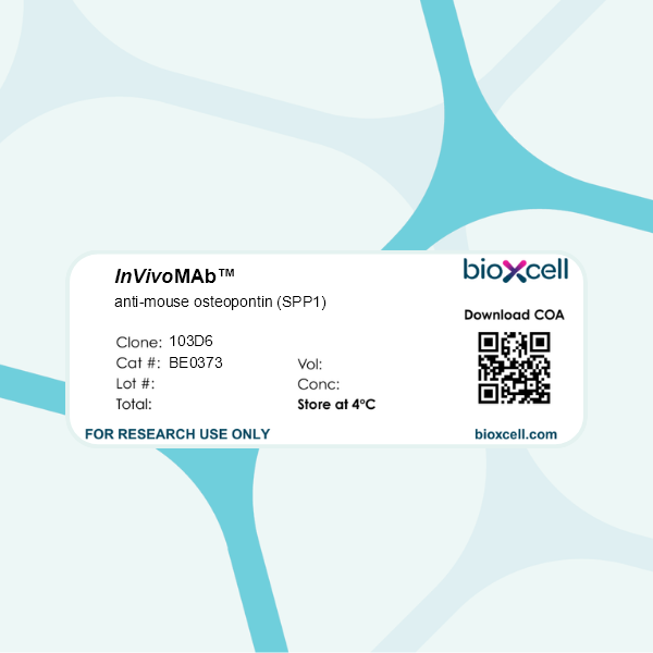
InVivoMAb anti-mouse osteopontin (SPP1)
Product Description
Specifications
| Isotype | Mouse IgG2c, κ |
|---|---|
| Recommended Isotype Control(s) | InVivoMAb mouse IgG2c isotype control, anti-dengue virus |
| Recommended Dilution Buffer | InVivoPure pH 7.0 Dilution Buffer |
| Conjugation | This product is unconjugated. Conjugation is available via our Antibody Conjugation Services. |
| Immunogen | Recombinant mouse OPN protein |
| Reported Applications |
in vivo OPN neutralization in vitro OPN neutralization ELISA |
| Formulation |
PBS, pH 7.0 Contains no stabilizers or preservatives |
| Endotoxin |
≤1EU/mg (≤0.001EU/μg) Determined by LAL assay |
| Purity |
≥95% Determined by SDS-PAGE |
| Sterility | 0.2 µm filtration |
| Purification | Protein G |
| RRID | AB_2927510 |
| Molecular Weight | 150 kDa |
| Storage | The antibody solution should be stored at the stock concentration at 4°C. Do not freeze. |
| Need a Custom Formulation? | See All Antibody Customization Options |
Application References
in vivo OPN neutralization
De Muynck K, Heyerick L, De Ponti FF, Vanderborght B, Meese T, Van Campenhout S, Baudonck L, Gijbels E, Rodrigues PM, Banales JM, Vesterhuus M, Folseraas T, Scott CL, Vinken M, Van der Linden M, Hoorens A, Van Dorpe J, Lefere S, Geerts A, Van Nieuwer
PubMed
Background and aims: Primary sclerosing cholangitis (PSC) is an immune-mediated cholestatic liver disease for which pharmacological treatment options are currently unavailable. PSC is strongly associated with colitis and a disruption of the gut-liver axis, and macrophages are involved in the pathogenesis of PSC. However, how gut-liver interactions and specific macrophage populations contribute to PSC is incompletely understood. Approach and results: We investigated the impact of cholestasis and colitis on the hepatic and colonic microenvironment, and performed an in-depth characterization of hepatic macrophage dynamics and function in models of concomitant cholangitis and colitis. Cholestasis-induced fibrosis was characterized by depletion of resident KCs, and enrichment of monocytes and monocyte-derived macrophages (MoMFs) in the liver. These MoMFs highly express triggering-receptor-expressed-on-myeloid-cells-2 ( Trem2 ) and osteopontin ( Spp1 ), markers assigned to hepatic bile duct-associated macrophages, and were enriched around the portal triad, which was confirmed in human PSC. Colitis induced monocyte/macrophage infiltration in the gut and liver, and enhanced cholestasis-induced MoMF- Trem2 and Spp1 upregulation, yet did not exacerbate liver fibrosis. Bone marrow chimeras showed that knockout of Spp1 in infiltrated MoMFs exacerbates inflammation in vivo and in vitro , while monoclonal antibody-mediated neutralization of SPP1 conferred protection in experimental PSC. In human PSC patients, serum osteopontin levels are elevated compared to control, and significantly increased in advanced stage PSC and might serve as a prognostic biomarker for liver transplant-free survival. Conclusions: Our data shed light on gut-liver axis perturbations and macrophage dynamics and function in PSC and highlight SPP1/OPN as a prognostic marker and future therapeutic target in PSC.
in vivo OPN neutralization
Bui TM, Yalom LK, Ning E, Urbanczyk JM, Ren X, Herrnreiter CJ, Disario JA, Wray B, Schipma MJ, Velichko YS, Sullivan DP, Abe K, Lauberth SM, Yang GY, Dulai PS, Hanauer SB, Sumagin R (2024). "Tissue-specific reprogramming leads to angiogenic neutrophi
PubMed
Neutrophil (PMN) tissue accumulation is an established feature of ulcerative colitis (UC) lesions and colorectal cancer (CRC). To assess the PMN phenotypic and functional diversification during the transition from inflammatory ulceration to CRC we analyzed the transcriptomic landscape of blood and tissue PMNs. Transcriptional programs effectively separated PMNs based on their proximity to peripheral blood, inflamed colon, and tumors. In silico pathway overrepresentation analysis, protein-network mapping, gene signature identification, and gene-ontology scoring revealed unique enrichment of angiogenic and vasculature development pathways in tumor-associated neutrophils (TANs). Functional studies utilizing ex vivo cultures, colitis-induced murine CRC, and patient-derived xenograft models demonstrated a critical role for TANs in promoting tumor vascularization. Spp1 (OPN) and Mmp14 (MT1-MMP) were identified by unbiased -omics and mechanistic studies to be highly induced in TANs, acting to critically regulate endothelial cell chemotaxis and branching. TCGA data set and clinical specimens confirmed enrichment of SPP1 and MMP14 in high-grade CRC but not in patients with UC. Pharmacological inhibition of TAN trafficking or MMP14 activity effectively reduced tumor vascular density, leading to CRC regression. Our findings demonstrate a niche-directed PMN functional specialization and identify TAN contributions to tumor vascularization, delineating what we believe to be a new therapeutic framework for CRC treatment focused on TAN angiogenic properties.
in vivo OPN neutralization
Bianchi E, Rontauroli S, Tavernari L, Mirabile M, Pedrazzi F, Genovese E, Sartini S, Dall', Ora M, Grisendi G, Fabbiani L, Maccaferri M, Carretta C, Parenti S, Fantini S, Bartalucci N, Calabresi L, Balliu M, Guglielmelli P, Potenza L, Tagliafico
PubMed
Clonal myeloproliferation and development of bone marrow (BM) fibrosis are the major pathogenetic events in myelofibrosis (MF). The identification of novel antifibrotic strategies is of utmost importance since the effectiveness of current therapies in reverting BM fibrosis is debated. We previously demonstrated that osteopontin (OPN) has a profibrotic role in MF by promoting mesenchymal stromal cells proliferation and collagen production. Moreover, increased plasma OPN correlated with higher BM fibrosis grade and inferior overall survival in MF patients. To understand whether OPN is a druggable target in MF, we assessed putative inhibitors of OPN expression in vitro and identified ERK1/2 as a major regulator of OPN production. Increased OPN plasma levels were associated with BM fibrosis development in the Romiplostim-induced MF mouse model. Moreover, ERK1/2 inhibition led to a remarkable reduction of OPN production and BM fibrosis in Romiplostim-treated mice. Strikingly, the antifibrotic effect of ERK1/2 inhibition can be mainly ascribed to the reduced OPN production since it could be recapitulated through the administration of anti-OPN neutralizing antibody. Our results demonstrate that OPN is a novel druggable target in MF and pave the way to antifibrotic therapies based on the inhibition of ERK1/2-driven OPN production or the neutralization of OPN activity.
in vivo OPN neutralization
in vitro OPN neutralization
ELISA
Klement, J. D., et al (2021). "Osteopontin Blockade Immunotherapy Increases Cytotoxic T Lymphocyte Lytic Activity and Suppresses Colon Tumor Progression" Cancers (Basel) 13(5).
PubMed
Human colorectal cancers are mostly microsatellite-stable with no response to anti-PD-1 blockade immunotherapy, necessitating the development of a new immunotherapy. Osteopontin (OPN) is elevated in human colorectal cancer and may function as an immune checkpoint. We aimed at elucidating the mechanism of action of OPN and determining the efficacy of OPN blockade immunotherapy in suppression of colon cancer. We report here that OPN is primarily expressed in tumor cells, myeloid cells, and innate lymphoid cells in human colorectal carcinoma. Spp1 knock out mice exhibit a high incidence and fast growth rate of carcinogen-induced tumors. Knocking out Spp1 in colon tumor cells increased tumor-specific CTL cytotoxicity in vitro and resulted in decreased tumor growth in vivo. The OPN protein level is elevated in the peripheral blood of tumor-bearing mice. We developed four OPN neutralization monoclonal antibodies based on their efficacy in blocking OPN inhibition of T cell activation. OPN clones 100D3 and 103D6 increased the efficacy of tumor-specific CTLs in killing colon tumor cells in vitro and suppressed colon tumor growth in tumor-bearing mice in vivo. Our data indicate that OPN blockade immunotherapy with 100D3 and 103D6 has great potential to be further developed for colorectal cancer immunotherapy and for rendering a colorectal cancer response to anti-PD-1 immunotherapy.
Product Citations
-
-
Mus musculus (Mouse)
-
Cancer Research
Inhibition of ERK1/2 signaling prevents bone marrow fibrosis by reducing osteopontin plasma levels in a myelofibrosis mouse model.
In Leukemia on 1 May 2023 by Bianchi, E., Rontauroli, S., et al.
PubMed
Clonal myeloproliferation and development of bone marrow (BM) fibrosis are the major pathogenetic events in myelofibrosis (MF). The identification of novel antifibrotic strategies is of utmost importance since the effectiveness of current therapies in reverting BM fibrosis is debated. We previously demonstrated that osteopontin (OPN) has a profibrotic role in MF by promoting mesenchymal stromal cells proliferation and collagen production. Moreover, increased plasma OPN correlated with higher BM fibrosis grade and inferior overall survival in MF patients. To understand whether OPN is a druggable target in MF, we assessed putative inhibitors of OPN expression in vitro and identified ERK1/2 as a major regulator of OPN production. Increased OPN plasma levels were associated with BM fibrosis development in the Romiplostim-induced MF mouse model. Moreover, ERK1/2 inhibition led to a remarkable reduction of OPN production and BM fibrosis in Romiplostim-treated mice. Strikingly, the antifibrotic effect of ERK1/2 inhibition can be mainly ascribed to the reduced OPN production since it could be recapitulated through the administration of anti-OPN neutralizing antibody. Our results demonstrate that OPN is a novel druggable target in MF and pave the way to antifibrotic therapies based on the inhibition of ERK1/2-driven OPN production or the neutralization of OPN activity.
-

