
InVivoMAb anti-mouse Ly6C
Flow cytometry
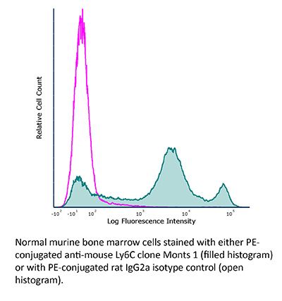
Product Description
Specifications
| Isotype | Rat IgG2a |
|---|---|
| Recommended Isotype Control(s) | InVivoMAb rat IgG2a isotype control, anti-trinitrophenol |
| Recommended Dilution Buffer | InVivoPure pH 7.0 Dilution Buffer |
| Conjugation | This product is unconjugated. Conjugation is available via our Antibody Conjugation Services. |
| Immunogen | Not available or unknown |
| Reported Applications |
in vivo macrophage depletion (in combination with clodronate liposomes) Flow cytometry |
| Formulation |
PBS, pH 7.0 Contains no stabilizers or preservatives |
| Endotoxin |
≤1EU/mg (≤0.001EU/μg) Determined by LAL assay |
| Purity |
≥95% Determined by SDS-PAGE |
| Sterility | 0.2 µm filtration |
| Production | Purified from cell culture supernatant in an animal-free facility |
| Purification | Protein G |
| RRID | AB_2687696 |
| Molecular Weight | 150 kDa |
| Storage | The antibody solution should be stored at the stock concentration at 4°C. Do not freeze. |
| Need a Custom Formulation? | See All Antibody Customization Options |
Application References
-
Rowe, A. M., et al (2017). "Subclinical Herpes Simplex Virus Type 1 Infections Provide Site-Specific Resistance to an Unrelated Pathogen" J Immunol. doi : 10.4049/jimmunol.1601310.
PubMed
HSV-1 infections of the cornea range in severity from minor transient discomfort to the blinding disease herpes stromal keratitis, yet most patients experience a single episode of epithelial keratitis followed by re-establishment of a clear cornea. We asked whether a single transient episode of HSV-1 epithelial keratitis causes long-term changes in the corneal microenvironment that influence immune responses to subsequent corneal infection or trauma. We showed that C57BL/6 mouse corneas infected with HSV-1 KOS, which induces transient herpes epithelial keratitis without herpes stromal keratitis sequelae, possessed a significant leukocytic infiltrate composed primarily of CD4+ T cells and macrophages along with elevated chemokines and cytokines that persisted without loss of corneal clarity (subclinical inflammation). Chemokine and cytokine expression was CD4+ T cell dependent, in that their production was significantly reduced by systemic CD4+ T cell depletion starting before infection, although short-term (3-d) local CD4+ T cell depletion postinfection did not influence chemokine levels in cornea. Corneas with subclinical inflammation developed significantly greater trauma-induced inflammation when they were recipients of syngeneic corneal transplants but also exhibited significantly increased resistance to infections by unrelated pathogens, such as pseudorabies virus. The resistance to pseudorabies virus was CD4+ T cell dependent, because it was eliminated by local CD4+ T cell depletion from the cornea. We conclude that transient HSV-1 corneal infections cause long-term alterations of the corneal microenvironment that provide CD4-dependent innate resistance to subsequent infections by antigenically unrelated pathogens.
-
del Rio, M. L., et al (2012). "Selective blockade of herpesvirus entry mediator-B and T lymphocyte attenuator pathway ameliorates acute graft-versus-host reaction" J Immunol 188(10): 4885-4896.
PubMed
The cosignaling network mediated by the herpesvirus entry mediator (HVEM; TNFRSF14) functions as a dual directional system that involves proinflammatory ligand, lymphotoxin that exhibits inducible expression and competes with HSV glycoprotein D for HVEM, a receptor expressed by T lymphocytes (LIGHT; TNFSF14), and the inhibitory Ig family member B and T lymphocyte attenuator (BTLA). To dissect the differential contributions of HVEM/BTLA and HVEM/LIGHT interactions, topographically-specific, competitive, and nonblocking anti-HVEM Abs that inhibit BTLA binding, but not LIGHT, were developed. We demonstrate that a BTLA-specific competitor attenuated the course of acute graft-versus-host reaction in a murine F(1) transfer semiallogeneic model. Selective HVEM/BTLA blockade did not inhibit donor T cell infiltration into graft-versus-host reaction target organs, but decreased the functional activity of the alloreactive T cells. These results highlight the critical role of HVEM/BTLA pathway in the control of the allogeneic immune response and identify a new therapeutic target for transplantation and autoimmune diseases.
-
Lubick, K., et al (2007). "Securinine, a GABAA receptor antagonist, enhances macrophage clearance of phase II C. burnetii: comparison with TLR agonists" J Leukoc Biol 82(5): 1062-1069.
PubMed
Innate immune cell stimulation represents a complementary approach to vaccines and antimicrobial drugs to counter infectious disease. We have used assays of macrophage activation and in vitro and in vivo phase II Coxiella burnetii infection models to compare and contrast the activity of a novel innate immune cell agonist, securinine, with known TLR agonists. As expected, TLR agonists, such as LPS (TLR4) and fibroblast-stimulating lipopeptide-1 (FSL-1; TLR2), induced macrophage activation and increased macrophage killing of phase II C. burnetii in vitro. FSL-1 also induced accelerated killing of C. burnetii in vivo. Securinine, a gamma-aminobutyric acid type A receptor antagonist, was found to induce TLR-independent macrophage activation in vitro, leading to IL-8 secretion, L-selectin down-regulation, and CD11b and MHC Class II antigen up-regulation. As seen with the TLR agonists, securinine also induced accelerated macrophage killing of C. burnetii in vitro and in vivo. In summary, as predicted by the literature, TLR agonists enhance macrophage killing of phase II C. burnetii in vitro, and at least for TLR2 agonists, this activity occurs in vivo as well. Securinine represents a novel macrophage agonist, which has similar effects as TLR agonists in this model yet apparently, does not act through known TLRs. Securinine has minimal toxicity in vivo, suggesting it or structurally similar compounds may represent novel, therapeutic adjuvants, which increase resistance to intracellular pathogens.
-
Duraiswamy, N., et al (1994). "Distinction of class II MHC+ Langerhans cell-like interstitial dendritic antigen-presenting cells in murine dermis from dermal macrophages" J Invest Dermatol 103(5): 678-683.
PubMed
Dermal cells are capable of initiating contact-hypersensitivity responses but the precise identification of the antigen-presenting cell within murine dermis is lacking. Class II major histocompatibility complex (MHC)+ cells with dendritic shape and lacking endothelial factor VIII but expressing the dendritic antigen-presenting cell marker NLDC-145 were observed in the perivascular and interstitial dermis of BALB/c and C3H/HeN skin. The heterogeneous class II MHC+ cells could be divided into two subsets: each was class II MHC+ CD45+ (bone marrow derived) GR-1- (non-neutrophil/macrophage) CD3- (non T), but one subset was CD11b+ (beta 2 integrin) and the other was CD11b-. Ultrastructural examination of class II MHC+ cells revealed the presence of a Langerhans cell-like/indeterminant cell subset with indented nuclei, dendritic morphology, active cytoplasm, and dense intermediate filaments. Phagolysomes and Birbeck granules were not observed in such cells, indicating these were distinct from dermal macrophages and from classical epidermal Langerhans cells, respectively. Cells with a monocyte/macrophage ultrastructural appearance were also noted, likely representing the class II MHC subset expressing CD11b and Ly6c (monocyte/endothelial antigen). Dermal cells in suspension were capable of processing and presenting large protein antigens to antigen-specific T-cell hybridomas; dermal cells also induced the syngeneic mixed lymphocyte reaction. The dermal antigen-presentation activities were totally abrogated by removal of class II MHC+ cells, but not by removal of CD11b+ cells or Ly6c+ cells, indicating that potent antigen-presenting cell activity was restricted to the class II MHC+ CD11b- Ly6c- subset (Langerhans cell-like/indeterminant cells). In conclusion, within a complex array of dermal leukocytes a murine dermal class II MHC+ cell population expressing a Langerhans cell-like/dendritic antigen-presenting cell phenotype and exhibiting potent antigen processing and presenting activity can be identified. The positioning of potent interstitial dendritic antigen-presenting cells at the interface of the vasculature with the dermal interstitium provides rapid access to an antigen-presenting cell as T cells first egress into the skin.
Product Citations
-
AARS1-mediated lactylation reprograms macrophage lipid metabolism to restrict antitumor immunity.
In J Immunother Cancer on 14 August 2026 by Xiao, Z., Meng, L., et al.
PubMed
Tumor-derived lactate has long been regarded as a metabolic waste product. However, accumulating evidence indicates that lactate also functions as a signaling molecule that actively remodels the tumor immune microenvironment. How lactate-driven post-translational modifications in immune cells contribute to immune evasion in hepatocellular carcinoma (HCC) remains incompletely understood. This study aimed to identify the immune cell population responsible for lactylation-driven immunosuppression in HCC and to elucidate the molecular mechanism by which lactylation rewires macrophage metabolism to impair CD8+ T cell-mediated antitumor immunity.
-
Tumoral extracellular vesicles and particles reprogram interstitial macrophages in the lung to promote vascular permeability and metastasis.
In Nat Cancer on 1 August 2026 by Dror, S., Wortzel, I., et al.
PubMed
The mechanisms by which tumor-derived extracellular vesicles and particles (EVPs) promote vascular permeability during premetastatic niche formation remain unclear. Here, we show that tumor EVPs rapidly induce vascular leakiness within 1 h of administration in female mice, creating a permissive environment that enhances metastatic seeding. Rather than acting directly on endothelial cells, EVPs activate NF-κB and JAK-STAT signaling in interstitial macrophages, leading to IL-6 secretion and increased vascular permeability. Interstitial macrophage depletion markedly reduces EVP-induced vascular leakiness and metastasis. We identify extracellular vesicle-associated integrin α5 (ITGα5) as a major functional determinant of this process, promoting macrophage activation and IL-6 secretion without affecting EVP uptake. EVPs derived from colorectal cancer tumors with high ITGα5 similarly induce macrophage IL-6 secretion and vascular permeability. Together, these findings define an EVP-macrophage-IL-6 axis that drives vascular permeability during premetastatic niche formation and identify EVP-associated ITGα5 as a key mediator of metastatic progression and a potential therapeutic target.
-
Dendritic cell redundancy enables priming of anti-tumor CD4+ T cells in pancreatic cancer.
In Cancer Cell on 13 July 2026 by Kureshi, C. T. S., Walsh, M. J., et al.
PubMed
Pancreatic ductal adenocarcinoma (PDAC) is resistant to current immunotherapies and lacks effective anti-tumor CD8+ T cells, which is potentially due to insufficient cross-presentation by cDC1s. Here, we combine a STING agonist with anti-CTLA-4 and anti-PD-1 to achieve durable remissions and immunologic memory in multiple mouse models of poorly immunogenic PDAC. We find that tumor control does not depend on CD8+ T cells or tumor cell MHC expression but instead requires IFNγ-producing CD4+ T cells (Th1s) that are primed by dendritic cells in lymph nodes. The triple combination immunotherapy induces an accumulation of activated cDC2s carrying tumor antigen into tumor-draining lymph nodes; cDC2s are required for orthotopic tumor clearance. Intratumoral CD4+ T cells and cDC2s remain present in treatment-naive and chemotherapy-exposed human PDAC. In chemotherapy-exposed patients' blood, cDC2s outnumber cDC1s by 10-fold. Therefore, therapeutic targeting of the cDC2-CD4+ T cell-IFNγ axis could be efficacious in PDAC.
-
IFNβ-pSTAT1-IRF1 axis controls colorectal tumor development through induction of immunogenic Ly6C+myeloid cells.
In Neoplasia on 1 January 2026 by Li, Y., Wu, Y., et al.
PubMed
It has been extensively studied that IFN-I inhibits tumor progression through their intrinsic functions in tumor cells, of which, regulating tumor cell immunogenicity is a critical way. Besides to tumor cells, myeloid cells also comprise a major component of tumor microenvironment. Nevertheless, whether IFN-I modulates the immunogenicity and function of myeloid cells in tumor microenvironment is less explored. In this research, we report that IFNβ effectively induces Ly6C+ myeloid cell differentiation in vitro in mouse. In human PBMCs, CD14+CD33+ monocytic population was consistently accumulated in the presence of IFNβ. In agreement, overexpression of IFNβ in tumor microenvironment by IFNBCOL01 treatment resulted in dramatic increase of tumor-infiltrating Ly6C+ myeloid cells and obvious tumor growth control in vivo. Of note, overexpression of IFNβ promotes the immunogenic Ly6C+CD103+CD11c+ subset accumulation in tumor-infiltrating myeloid cells. At the molecular level, we illustrated that the pSTAT1 directly binds to the irf1 promoter and that IRF1 directly binds to the Ly6C promoter in myeloid cells both in vitro and in vivo. Furthermore, anti-Ly6C blockade therapy significantly reversed the anti-tumor effect of IFNBCOL01 by restoring NOS2 expression and consequently suppressing T cell functions in tumor-bearing mice. Overall, our findings determine that IFNβ drives myeloid cells to differentiate into Ly6C+ subset via the stimulation of pSTAT1-IRF1 axis in myeloid cells. In addition, IFNβ inhibits the immunosuppressive enzyme NOS2 expression in Ly6C+ myeloid cells, which may decrease the potential immunosuppressive function of NOS2 to boost the T cell function to repress tumor, thus proposing a new perspective for the anti-tumor mechanism of IFN-I.