InVivoMAb anti-mouse CSF1R (CD115)
Product Description
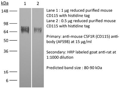
Specifications
| Isotype | Rat IgG2a, κ |
|---|---|
| Recommended Isotype Control(s) | InVivoMAb rat IgG2a isotype control, anti-trinitrophenol |
| Recommended Dilution Buffer | InVivoPure pH 7.0 Dilution Buffer |
| Conjugation | This product is unconjugated. Conjugation is available via our Antibody Conjugation Services. |
| Immunogen | Not available or unknown |
| Reported Applications |
in vivo macrophage depletion in vitro CSF1R neutralization in vivo monocyte depletion Flow cytometry Western blot |
| Formulation |
PBS, pH 7.0 Contains no stabilizers or preservatives |
| Endotoxin |
≤1EU/mg (≤0.001EU/μg) Determined by LAL assay |
| Purity |
≥95% Determined by SDS-PAGE |
| Sterility | 0.2 µm filtration |
| Production | Purified from cell culture supernatant in an animal-free facility |
| Purification | Protein G |
| RRID | AB_2687699 |
| Molecular Weight | 150 kDa |
| Storage | The antibody solution should be stored at the stock concentration at 4°C. Do not freeze. |
| Need a Custom Formulation? | See All Antibody Customization Options |
Application References
-
Bauche, D., et al (2018). "LAG3(+) Regulatory T Cells Restrain Interleukin-23-Producing CX3CR1(+) Gut-Resident Macrophages during Group 3 Innate Lymphoid Cell-Driven Colitis" Immunity 49(2): 342-352 e345.
PubMed
Interleukin-22 (IL-22)-producing group 3 innate lymphoid cells (ILC3) maintains gut homeostasis but can also promote inflammatory bowel disease (IBD). The regulation of ILC3-dependent colitis remains to be elucidated. Here we show that Foxp3(+) regulatory T cells (Treg cells) prevented ILC3-mediated colitis in an IL-10-independent manner. Treg cells inhibited IL-23 and IL-1beta production from intestinal-resident CX3CR1(+) macrophages but not CD103(+) dendritic cells. Moreover, Treg cells restrained ILC3 production of IL-22 through suppression of CX3CR1(+) macrophage production of IL-23 and IL-1beta. This suppression was contact dependent and was mediated by latent activation gene-3 (LAG-3)-an immune checkpoint receptor-expressed on Treg cells. Engagement of LAG-3 on MHC class II drove profound immunosuppression of CX3CR1(+) tissue-resident macrophages. Our study reveals that the health of the intestinal mucosa is maintained by an axis driven by Treg cells communication with resident macrophages that withhold inflammatory stimuli required for ILC3 function.
-
Gordon, S. R., et al (2017). "PD-1 expression by tumour-associated macrophages inhibits phagocytosis and tumour immunity" Nature 545(7655): 495-499.
PubMed
Programmed cell death protein 1 (PD-1) is an immune checkpoint receptor that is upregulated on activated T cells for the induction of immune tolerance. Tumour cells frequently overexpress the ligand for PD-1, programmed cell death ligand 1 (PD-L1), facilitating their escape from the immune system. Monoclonal antibodies that block the interaction between PD-1 and PD-L1, by binding to either the ligand or receptor, have shown notable clinical efficacy in patients with a variety of cancers, including melanoma, colorectal cancer, non-small-cell lung cancer and Hodgkin’s lymphoma. Although it is well established that PD-1-PD-L1 blockade activates T cells, little is known about the role that this pathway may have in tumour-associated macrophages (TAMs). Here we show that both mouse and human TAMs express PD-1. TAM PD-1 expression increases over time in mouse models of cancer and with increasing disease stage in primary human cancers. TAM PD-1 expression correlates negatively with phagocytic potency against tumour cells, and blockade of PD-1-PD-L1 in vivo increases macrophage phagocytosis, reduces tumour growth and lengthens the survival of mice in mouse models of cancer in a macrophage-dependent fashion. This suggests that PD-1-PD-L1 therapies may also function through a direct effect on macrophages, with substantial implications for the treatment of cancer with these agents.
-
Moynihan, K. D., et al (2016). "Eradication of large established tumors in mice by combination immunotherapy that engages innate and adaptive immune responses" Nat Med. doi : 10.1038/nm.4200.
PubMed
Checkpoint blockade with antibodies specific for cytotoxic T lymphocyte-associated protein (CTLA)-4 or programmed cell death 1 (PDCD1; also known as PD-1) elicits durable tumor regression in metastatic cancer, but these dramatic responses are confined to a minority of patients. This suboptimal outcome is probably due in part to the complex network of immunosuppressive pathways present in advanced tumors, which are unlikely to be overcome by intervention at a single signaling checkpoint. Here we describe a combination immunotherapy that recruits a variety of innate and adaptive immune cells to eliminate large tumor burdens in syngeneic tumor models and a genetically engineered mouse model of melanoma; to our knowledge tumors of this size have not previously been curable by treatments relying on endogenous immunity. Maximal antitumor efficacy required four components: a tumor-antigen-targeting antibody, a recombinant interleukin-2 with an extended half-life, anti-PD-1 and a powerful T cell vaccine. Depletion experiments revealed that CD8+ T cells, cross-presenting dendritic cells and several other innate immune cell subsets were required for tumor regression. Effective treatment induced infiltration of immune cells and production of inflammatory cytokines in the tumor, enhanced antibody-mediated tumor antigen uptake and promoted antigen spreading. These results demonstrate the capacity of an elicited endogenous immune response to destroy large, established tumors and elucidate essential characteristics of combination immunotherapies that are capable of curing a majority of tumors in experimental settings typically viewed as intractable.
-
Arnold, I. C., et al (2015). "CD11c monocyte/macrophages promote chronic Helicobacter hepaticus-induced intestinal inflammation through the production of IL-23" Mucosal Immunol. doi : 10.1038/mi.2015.65.
PubMed
In inflammatory bowel diseases, a breakdown in host microbial interactions accompanies sustained activation of immune cells in the gut. Functional studies suggest a key role for interleukin-23 (IL-23) in orchestrating intestinal inflammation. IL-23 can be produced by various mononuclear phagocytes (MNPs) following acute microbial stimulation, but little is known about the key cellular sources of IL-23 that drive chronic intestinal inflammation. Here we have addressed this question using a physiological model of bacteria-driven colitis. By combining conditional gene ablation and gene expression profiling, we found that IL-23 production by CD11c+ MNPs was essential to trigger intestinal immunopathology and identified MHCII+ monocytes and macrophages as the major source of IL-23. Expression of IL-23 by monocytes was acquired during their differentiation in the intestine and correlated with the expression of major histocompatibility complex class II (MHCII) and CD64. In contrast, Batf3-dependent CD103+ CD11b- dendritic cells were dispensable for bacteria-induced colitis in this model. These studies reinforce the pathogenic role of monocytes in dysregulated responses to intestinal bacteria and identify production of IL-23 as a key component of this response. Further understanding of the functional sources of IL-23 in diverse forms of intestinal inflammation may lead to novel therapeutic strategies aimed at interrupting IL-23-driven immune pathology.Mucosal Immunology advance online publication 5 August 2015. doi:10.1038/mi.2015.65.
Product Citations
-
Tumor-derived branched-chain α-keto acids activate Notch signaling in tumor-associated macrophages to limit immunity.
In Nat Immunol on 1 June 2026 by Ma, Q. X., Zhao, R., et al.
PubMed
Tumor cells are highly dependent on branched-chain amino acids, which can activate mechanistic target of rapamycin complex 1, but the downstream catabolite branched-chain α-keto acids (BCKAs) are not well studied in this context. Here, using clinical samples and genetically engineered mouse tumor models, we showed that tumor-derived BCKAs are secreted actively into the tumor microenvironment (TME) where they reprogram tumor-associated macrophages (TAMs) to promote tumor progression. Through genome-wide CRISPR screening, we identified Notch2 as a direct molecular target of BCKAs. BCKAs activate Notch signaling by binding to and stabilizing cleaved Notch2, functionally reprogramming TAMs and fostering an immunosuppressive TME. Mutation of the BCKA-binding site in Notch2 abolishes this effect in vivo. Together, these findings identify BCKAs as signaling metabolites that mediate tumor immunosuppression through direct sensing by Notch2.
-
Tumor-immune-neural circuit disrupts energy homeostasis in cancer cachexia.
In Cancer Cell on 11 May 2026 by Shi, X., Arreola, A. X., et al.
PubMed
Cancer-induced cachexia and anorexia are debilitating complications across many cancers, yet effective treatments remain limited due to a poor understanding of the underlying mechanisms. Here, we identify an uncharacterized tumor-immune-neural circuit driving these syndromes, centered on growth and differentiation factor 15 (GDF15). Using genetically engineered mouse models, we find that loss of GDF15 protects against appetite loss, muscle wasting, and fat loss in pancreatic, lung, and skin cancers. Single-cell RNA sequencing reveals macrophages as a major source of GDF15, induced by tumor-derived colony-stimulating factor 1 (CSF1). GDF15 acts via the central nervous system to enhance β-adrenergic signaling in the tumor microenvironment, thereby amplifying cachexia. The disruption of this feedforward loop with GDF15-neutralizing antibody, anti-CSF1R antibody, or Rearranged during Transfection (RET) inhibitor markedly reduces both cachexia and anorexia. These findings reveal a non-cell-autonomous mechanism linking tumor signals, macrophage-derived GDF15, and neural pathways, highlighting the tumor-immune-neural triad as a promising therapeutic target.
-
The liver regulates ectopic calcification in Abcc6-deficient models of pseudoxanthoma elasticum.
In J Clin Invest on 1 May 2026 by Wang, Y., Sun, B., et al.
PubMed
Pseudoxanthoma Elasticum (PXE) is a rare disease caused by loss of function of the ATP-binding cassette C (ABC) member 6 (Abcc6) gene and characterized by ectopic calcification of multiple tissues, but the physiological reasons underlying ectopic calcification in PXE remain unclear. In a murine model of Abcc6-deficient PXE in which animals developed robust cardiac calcification after heart injury, we show the critical importance of the liver in mediating ectopic cardiac calcification. Tissue-specific deletion of Abcc6 in the liver, but not in the heart, was sufficient to cause post-injury cardiac calcification. Metabolomics and gene expression analysis demonstrated deficiencies in nucleotide metabolism, cellular energetics, and defects in cellular respiration underlying ectopic calcification in PXE. Functional abnormalities in cellular respiration in the injured heart were similar in animals with global or liver-specific Abcc6 deficiency, showing that hepatic Abcc6 expression regulated cellular respiration in the injured heart. We show that ectopic calcification in PXE was primarily dystrophic and that treatment with clodronate or etidronate, which prevent the growth of calcium hydroxyapatite mineralization, was sufficient to rescue the phenotype of ectopic cardiac calcification in Abcc6-deficient states. Taken together, these observations highlight the role of the liver in regulating target tissue metabolic and mitochondrial function in causing ectopic calcification in Abcc6-deficient states.
-
Immunogenic tumor cell death and T-cell-derived IFN-γ elicit tumoricidal macrophages to potentiate OX40 immunotherapy.
In Cell Rep Med on 21 April 2026 by Liu, Y., Zhao, J., et al.
PubMed
Understanding the mechanisms limiting OX40 agonist antibody efficacy is critical for developing more effective combination immunotherapies. Tumor microenvironment (TME) analysis revealed that OX40-antibody-responsive mice harbored tumor-associated macrophages (TAMs) with elevated NOS2 expression and heightened pattern recognition receptor (PRR) activation and interferon gamma (IFN-γ) signaling. In addition, patients with more favorable treatment responses to OX40 antibody therapy exhibited increased NOS2 expression. Mechanistically, tumor-infiltrating T-cell-derived IFN-γ synergizes with endogenous ligands of PRR released during immunogenic cell death to drive NOS2+ TAMs reprogramming. Translating these insights into therapeutic strategy, a Combo approach composing of MPLA, IFN-γ, and OX40 agonist antibody is designed to actively polarize TAMs to express NOS2, which mediate tumor clearance through an NOS2-dependent cytotoxicity. Moreover, OX40-antibody-mediated regulatory T cell (Treg) depletion potentiated NOS2+ macrophage induction. This multimodal strategy offers a promising solution to overcome the limitations of OX40 antibody monotherapy and enhance outcomes of the OX40-targeted immunotherapies.