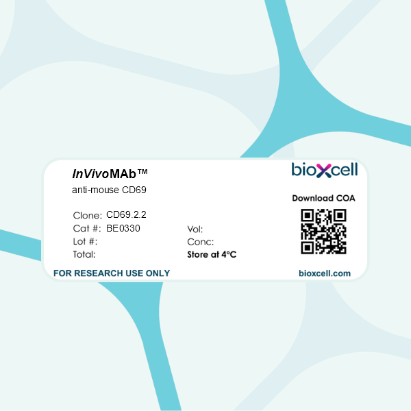
InVivoMAb anti-mouse CD69
Product Description
Specifications
| Isotype | Mouse IgG1, κ |
|---|---|
| Recommended Isotype Control(s) | InVivoMAb mouse IgG1 isotype control, unknown specificity |
| Recommended Dilution Buffer | InVivoPure pH 7.0 Dilution Buffer |
| Conjugation | This product is unconjugated. Conjugation is available via our Antibody Conjugation Services. |
| Immunogen | CD69+ murine 300-19 pre-B cells |
| Reported Applications |
in vivo down-regulation of CD69 expression Functional assays |
| Formulation |
PBS, pH 7.0 Contains no stabilizers or preservatives |
| Endotoxin |
≤1EU/mg (≤0.001EU/μg) Determined by LAL assay |
| Purity |
≥95% Determined by SDS-PAGE |
| Sterility | 0.2 µm filtration |
| Production | Purified from cell culture supernatant in an animal-free facility |
| Purification | Protein A |
| RRID | AB_2894750 |
| Molecular Weight | 150 kDa |
| Storage | The antibody solution should be stored at the stock concentration at 4°C. Do not freeze. |
| Need a Custom Formulation? | See All Antibody Customization Options |
Application References
in vivo down-regulation of CD69 expression
Notario, L., et al (2019). "CD69 Targeting Enhances Anti-vaccinia Virus Immunity" J Virol 93(19).
PubMed
CD69 is highly expressed on the leukocyte surface upon viral infection, and its regulatory role in the vaccinia virus (VACV) immune response has been recently demonstrated using CD69(-/-) mice. Here, we show augmented control of VACV infection using the anti-human CD69 monoclonal antibody (MAb) 2.8 as both preventive and therapeutic treatment for mice expressing human CD69. This control was related to increased natural killer (NK) cell reactivity and increased numbers of cytokine-producing T and NK cells in the periphery. Moreover, similarly increased immunity and protection against VACV were reproduced over both long and short periods in anti-mouse CD69 MAb 2.2-treated immunocompetent wild-type (WT) mice and immunodeficient Rag2(-/-) CD69(+/+) mice. This result was not due to synergy between infection and anti-CD69 treatment since, in the absence of infection, anti-human CD69 targeting induced immune activation, which was characterized by mobilization, proliferation, and enhanced survival of immune cells as well as marked production of several innate proinflammatory cytokines by immune cells. Additionally, we showed that the rapid leukocyte effect induced by anti-CD69 MAb treatment was dependent on mTOR signaling. These properties suggest the potential of CD69-targeted therapy as an antiviral adjuvant to prevent derived infections.IMPORTANCE In this study, we demonstrate the influence of human and mouse anti-CD69 therapies on the immune response to VACV infection. We report that targeting CD69 increases the leukocyte numbers in the secondary lymphoid organs during infection and improves the capacity to clear the viral infection. Targeting CD69 increases the numbers of gamma interferon (IFN-gamma)- and tumor necrosis factor alpha (TNF-alpha)-producing NK and T cells. In mice expressing human CD69, treatment with an anti-CD69 MAb produces increases in cytokine production, survival, and proliferation mediated in part by mTOR signaling. These results, together with the fact that we have mainly worked with a human-CD69 transgenic model, reveal CD69 as a treatment target to enhance vaccine protectiveness.
Functional Assays
Cortes, J. R., et al (2014). "Maintenance of immune tolerance by Foxp3+ regulatory T cells requires CD69 expression" J Autoimmun 55: 51-62.
PubMed
Although FoxP3(+) regulatory T cells are key players in the maintenance of immune tolerance and autoimmunity, the lack of specific markers constitute an obstacle to their use for immunotherapy protocols. In this study, we have investigated the role of the C-type lectin receptor CD69 in the suppressor function of Tregs and maintenance of immune tolerance towards harmless inhaled antigens. We identified a novel FoxP3(+)CD69(+) Treg subset capable to maintain immune tolerance and protect to developing inflammation. Although CD69(+) and CD69(-)FoxP3(+) Tregs exist in homeostasis, only CD69-expressing Tregs express high levels of CTLA-4, ICOS, CD38 and GITR suppression-associated markers, secrete high amounts of TGFbeta and have potent suppressor activity. This activity is regulated by STAT5 and ERK signaling pathways and is impaired by antibody-mediated down-regulation of CD69 expression. Moreover, immunotherapy with FoxP3(+)CD69(+) Tregs restores the homeostasis in Cd69(-/-) mice, that fail to induce tolerance, and is also highly proficient in the prevention of inflammation. The identification of the FoxP3(+)CD69(+) Treg subset paves the way toward the development of new therapeutic strategies to control immune homeostasis and autoimmunity.
in vivo down-regulation of CD69 expression
Lamana, A., et al (2006). "The role of CD69 in acute neutrophil-mediated inflammation" Eur J Immunol 36(10): 2632-2638.
PubMed
The leukocyte activation marker CD69 functions as a negative regulator of the immune response, both in NK-dependent tumor rejection and in the inflammation associated with lymphocyte-dependent collagen-induced arthritis. In contrast, it has been reported that CD69-deficient mice are refractory to the neutrophil-dependent acute inflammatory response associated with anti-type II collagen antibody-induced arthritis (CAIA), suggesting a positive regulatory role for CD69 in neutrophil function during arthritis induction. To clarify this discrepancy, the CAIA response was independently analyzed in our CD69-deficient mice. In these experiments, the inflammatory response was unaffected by CD69 deficiency. Additionally, the in vivo down-regulation of CD69 expression by treatment of wild-type mice with the anti-CD69 mAb 2.2, which mimics the CD69-deficient phenotype, did not affect the course of arthritis in this model. Moreover, down-regulation of CD69 expression increased expression in arthritic joints of key inflammatory mediators, including IL-1beta, IL-6 and the chemokine MCP-1. Neutrophil accumulation in zymosan-treated air pouches and in thioglycolate-treated peritoneal cavities was also unaffected in CD69-deficient mice. In addition, CD69 expression was absent in activated neutrophils. Taken together, these results rule out a significant stimulatory role for CD69 in acute inflammatory responses mediated by neutrophils.
in vivo down-regulation of CD69 expression
Functional Assays
Esplugues, E., et al (2005). "Induction of tumor NK-cell immunity by anti-CD69 antibody therapy" Blood 105(11): 4399-4406.
PubMed
The leukocyte activation marker CD69 is a novel regulator of the immune response, modulating the production of cytokines including transforming growth factor-beta (TGF-beta). We have generated an antimurine CD69 monoclonal antibody (mAb), CD69.2.2, which down-regulates CD69 expression in vivo but does not deplete CD69-expressing cells. Therapeutic administration of CD69.2.2 to wild-type mice induces significant natural killer (NK) cell-dependent antitumor responses to major histocompatibility complex (MHC) class I low RMA-S lymphomas and to RM-1 prostatic carcinoma lung metastases. These in vivo antitumor responses are comparable to those seen in CD69(-/-) mice. Enhanced host NK cytotoxic activity correlates with a reduction in NK-cell TGF-beta production and is independent of tumor priming. In vitro studies demonstrate the novel ability of anti-CD69 mAbs to activate resting NK cells in an Fc receptor-independent manner, resulting in a substantial increase in both NK-cell cytolytic activity and interferon gamma (IFNgamma) production. Modulation of the innate immune system with monoclonal antibodies to host CD69 thus provides a novel means to antagonize tumor growth and metastasis.
in vivo down-regulation of CD69 expression
Esplugues, E., et al (2003). "Enhanced antitumor immunity in mice deficient in CD69" J Exp Med 197(9): 1093-1106.
PubMed
We investigated the in vivo role of CD69 by analyzing the susceptibility of CD69-/- mice to tumors. CD69-/- mice challenged with MHC class I- tumors (RMA-S and RM-1) showed greatly reduced tumor growth and prolonged survival compared with wild-type (WT) mice. The enhanced anti-tumor response was NK cell and T lymphocyte-mediated, and was due, at least in part, to an increase in local lymphocytes. Resistance of CD69-/- mice to MHC class I- tumor growth was also associated with increased production of the chemokine MCP-1, diminished TGF-beta production, and decreased lymphocyte apoptosis. Moreover, the in vivo blockade of TGF-beta in WT mice resulted in enhanced anti-tumor response. In addition, CD69 engagement induced NK and T cell production of TGF-beta, directly linking CD69 signaling to TGF-beta regulation. Furthermore, anti-CD69 antibody treatment in WT mice induced a specific down-regulation in CD69 expression that resulted in augmented anti-tumor response. These data unmask a novel role for CD69 as a negative regulator of anti-tumor responses and show the possibility of a novel approach for the therapy of tumors.
Product Citations
-
-
Mus musculus (Mouse)
-
Immunology and Microbiology
Pathobiont-induced suppressive immune imprints thwart T cell vaccine responses.
In Nat Commun on 16 December 2024 by Hajam, I. A., Tsai, C. M., et al.
PubMed
Pathobionts have evolved many strategies to coexist with the host, but how immune evasion mechanisms contribute to the difficulty of developing vaccines against pathobionts is unclear. Meanwhile, Staphylococcus aureus (SA) has resisted human vaccine development to date. Here we show that prior SA exposure induces non-protective CD4+ T cell imprints, leading to the blunting of protective IsdB vaccine responses. Mechanistically, these SA-experienced CD4+ T cells express IL-10, which is further amplified by vaccination and impedes vaccine protection by binding with IL-10Rα on CD4+ T cell and inhibit IL-17A production. IL-10 also mediates cross-suppression of IsdB and sdrE multi-antigen vaccine. By contrast, the inefficiency of SA IsdB, IsdA and MntC vaccines can be overcome by co-treatment with adjuvants that promote IL-17A and IFN-γ responses. We thus propose that IL-10 secreting, SA-experienced CD4+ T cell imprints represent a staphylococcal immune escaping mechanism that needs to be taken into consideration for future vaccine development.
-

