InVivoMAb anti-mouse CD45RB
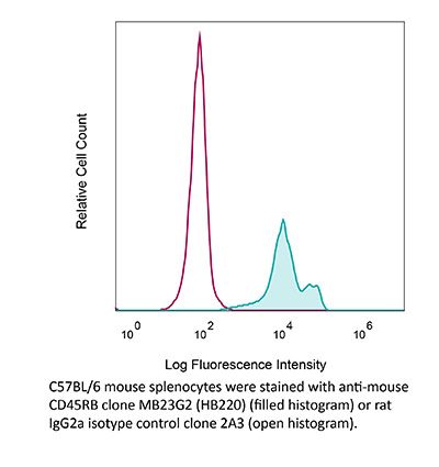
Product Description
Specifications
| Isotype | Rat IgG2a, κ |
|---|---|
| Recommended Isotype Control(s) | InVivoMAb rat IgG2a isotype control, anti-trinitrophenol |
| Recommended Dilution Buffer | InVivoPure pH 7.0 Dilution Buffer |
| Conjugation | This product is unconjugated. Conjugation is available via our Antibody Conjugation Services. |
| Immunogen | Anti-immunoglobulin activated mouse B cells |
| Reported Applications |
in vivo anti-CD45RB_mediated tolerance induction in vivo pre-mNK cell depletion |
| Formulation |
PBS, pH 7.0 Contains no stabilizers or preservatives |
| Endotoxin |
≤1EU/mg (≤0.001EU/μg) Determined by LAL assay |
| Purity |
≥95% Determined by SDS-PAGE |
| Sterility | 0.2 µm filtration |
| Production | Purified from cell culture supernatant in an animal-free facility |
| Purification | Protein G |
| RRID | AB_1107653 |
| Molecular Weight | 150 kDa |
| Storage | The antibody solution should be stored at the stock concentration at 4°C. Do not freeze. |
| Need a Custom Formulation? | See All Antibody Customization Options |
Application References
-
Wilson, K. A., et al (2015). "Depletion of B220NK1.1 cells enhances the rejection of established melanoma by tumor-specific CD4 T cells" Oncoimmunology 4(8): e1019196.
PubMed
Five-year survival rates for patients diagnosed with metastatic melanoma are less than 5%. Adoptive cell transfer (ACT) has achieved an objective response of 50% by Response Evaluation Criteria in Solid Tumors (RECIST) in this patient population. For ACT to be maximally effective, the host must first be lymphodepleted. It is hypothesized that lymphodepletion may remove regulatory elements and cytokine sinks, or increase the activation and availability of antigen presenting cells (APCs). We use an in vivo model to study the ACT of tumor-associated antigen (TAA)-specific CD4+ T cells (TRP-1 cells). We have discovered that depletion of NK1.1+ cells enhances the rejection of established melanoma tumors by adoptively transferred TRP-1 CD4+ T cells. NK1.1+ cell depletion increases the number of CD4+ T cells, the serum concentration of pro-inflammatory cytokines, autoimmune vitiligo, host survival and prevented recurrence after ACT. Because multiple cells express NK1.1, we targeted different NK1.1+ cell populations using antibodies specific for NK cells, pre-mNK cells, and innate lymphoid cells (ILCs). Our data suggests that NK1.1+B220+ pre-mNK cells (also known as interferon-producing killer dendritic cells; IKDCs) are an important inhibitor of the CD4+ T cell response to melanoma. Understanding this mechanism may help design new immunotherapies to modulate the activity of pre-mNKs in the face of an antitumor immune response and inhibit their suppression of adoptively transferred T cells.
-
Shen, H. and D. R. Goldstein (2009). "IL-6 and TNF-alpha synergistically inhibit allograft acceptance" J Am Soc Nephrol 20(5): 1032-1040.
PubMed
Previous studies suggested that activation of the innate immune system impairs the induction of transplantation tolerance, but the responsible inflammatory mediators have not been identified. In this study, we examined whether IL-6 and TNF-alpha promote resistance to transplantation tolerance. Using a highly immunogenic murine skin allograft model, we found that the absence of both IL-6 and TNF-alpha in the graft recipient synergized with co-stimulatory blockade to induce tolerance. Furthermore, IL-6 and TNF-alpha acted together to promote T cell alloimmune responses both in vitro and in vivo and to impair the ability of regulatory T cells to suppress effector T cell alloimmunity. In addition, deficiency of recipient IRAK-M, a negative regulator of certain innate immune pathways, augmented cellular IL-6 and TNF-alpha responses and impaired the ability of co-stimulatory blockade to extend allograft survival. In summary, IL-6 and TNF-alpha synergistically impair the efficacy of therapies that promote allograft acceptance.
-
Lee, K. M., et al (2014). "TGF-beta-producing regulatory B cells induce regulatory T cells and promote transplantation tolerance" Eur J Immunol 44(6): 1728-1736.
PubMed
Regulatory B (Breg) cells have been shown to play a critical role in immune homeostasis and in autoimmunity models. We have recently demonstrated that combined anti-T cell immunoglobulin domain and mucin domain-1 and anti-CD45RB antibody treatment results in tolerance to full MHC-mismatched islet allografts in mice by generating Breg cells that are necessary for tolerance. Breg cells are antigen-specific and are capable of transferring tolerance to untreated, transplanted animals. Here, we demonstrate that adoptively transferred Breg cells require the presence of regulatory T (Treg) cells to establish tolerance, and that adoptive transfer of Breg cells increases the number of Treg cells. Interaction with Breg cells in vivo induces significantly more Foxp3 expression in CD4(+) CD25(-) T cells than with naive B cells. We also show that Breg cells express the TGF-beta associated latency-associated peptide and that Breg-cell mediated graft prolongation post-adoptive transfer is abrogated by neutralization of TGF-beta activity. Breg cells, like Treg cells, demonstrate preferential expression of both C-C chemokine receptor 6 and CXCR3. Collectively, these findings suggest that in this model of antibody-induced transplantation tolerance, Breg cells promote graft survival by promoting Treg-cell development, possibly via TGF-beta production.
-
Stocks, B. T., et al (2015). "Lupus-Prone Mice Resist Immune Regulation and Transplant Tolerance Induction" Am J Transplant .
PubMed
The strongly immunogenic environment in autoimmune diseases such as lupus may pose a stringent barrier to transplantation. Despite available murine models of lupus, transplant tolerance in this setting has yet to be fully investigated in highly penetrant genetic models of disease. Such studies are of clear clinical importance because lupus is a transplant indication in which transplanted kidneys have a substantially increased risk of rejection including a role for recurrent nephritis. In the fully penetrant B6.SLE123 mouse, we determined that CD4 T follicular helper and germinal center B cells were significantly expanded compared with healthy controls. We traced this expansion to resistance of effector CD4 T and B cells in B6.SLE123 mice to regulation by either CD4 T regulatory cells (CD4Tregs) or CD8 T regulatory cells (CD8Tregs), despite demonstrating normal function by Tregs in this strain. Finally, we determined that B6.SLE123 mice resist anti-CD45RB-mediated tolerance induction to foreign islet allografts, even in the absence of islet autoimmunity. Overall, B6.SLE123 lupus-prone mice are highly resistant to transplant tolerance induction, which provides a new model of failed tolerance in autoimmunity that may elucidate barriers to clinical transplantation in lupus through further cellular and genetic dissection.
Product Citations
-
Macrophages rescue cells from ferroptotic death.
In Cell Death Dis on 1 December 2025 by Hefetz, R., Lianski, S., et al.
PubMed
Ferroptosis, a non-apoptotic form of cell death marked by iron-dependent lipid peroxidation, has a key role in organ injury, degenerative disease, and vulnerability of therapy-resistant cancers. Although substantial progress has been made in understanding the molecular processes relevant to ferroptosis, additional cell-extrinsic processes that determine cell sensitivity toward ferroptosis remain unknown. Here we demonstrate that macrophages co-cultured with ferroptotic cancer cells from various types effectively mitigate cell death induced by GPX4 inhibitors (RSL3 and ML162), GPX4 silencing via shRNA, or the Xc- system inhibitor IKE. Furthermore, macrophages effectively reduced lipid peroxidation in ferroptotic cells. Importantly, macrophage function relies on direct cell-to-cell contact and is affected by their differentiation. Specifically, polarization into M1 macrophages, but not M2, greatly hinders their protective capabilities. Interestingly, unlike apoptotic cells, ferroptotic cells retain elevated levels of the 'don't eat me' signal, CD47, and conversely, fail to present the "eat me" signal phosphatidylserine (PS) on the outer layer of the plasma membrane, providing an opportunity for their rescue. Furthermore, in placental villi explants, macrophages protect trophoblasts from ferroptotic death. These results underscore the intricate interplay between ferroptotic cells and their microenvironment and provide compelling evidence of a yet-unrecognized anti-ferroptotic activity of macrophages as a cell-extrinsic mechanism that could be exploited by cancer cells to escape ferroptosis.
-
Protocol for renal subcapsular islet transplantation in diabetic mice to induce long-term immune tolerance.
In STAR Protoc on 20 June 2025 by Yuan, Y., Wu, Q., et al.
PubMed
Murine renal subcapsular islet transplantation presents a promising technique for diabetes treatment by addressing challenges such as immune rejection and reliance on immunosuppressive drugs. Here, we present a protocol for the isolation, purification, and transplantation of mouse pancreatic islets that overcomes these challenges. Specifically, we describe steps for inducing diabetes with streptozotocin, pancreatic perfusion and isolation, and islet cell purification. We then detail procedures for renal subcapsular islet transplantation, dual antibody therapy, and immune cell and graft monitoring. For complete details on the use and execution of this protocol, please refer to Liu et al.1.
-
Versatile tissue-injectable hydrogels capable of the extended hydrolytic release of bioactive protein therapeutics.
In Bioeng Transl Med on 1 September 2024 by Nealy, E. S., Reed, J., et al.
PubMed
Hydrogels are extensively employed in healthcare due to their adaptable structures, high water content, and biocompatibility, with FDA-approved applications ranging from spinal cord regeneration to local therapeutic delivery. However, clinical hydrogels encounter challenges related to inconsistent therapeutic exposure, unmodifiable release windows, and difficulties in subsurface polymer insertion. Addressing these issues, we engineered injectable, biocompatible hydrogels as a local therapeutic depot, utilizing poly(ethylene glycol) (PEG)-based hydrogels functionalized with bioorthogonal SPAAC handles for network polymerization and functionalization. Our hydrogel solutions polymerize in situ in a temperature-sensitive manner, persist in tissue, and facilitate the delivery of bioactive therapeutics in subsurface locations. Demonstrating the efficacy of our approach, recombinant anti-CD47 monoclonal antibodies, when incorporated into subsurface-injected hydrogel solutions, exhibited cytotoxic activity against infiltrative high-grade glioma xenografts in the rodent brain. To enhance the gel's versatility, recombinant protein cargos can undergo site-specific modification with hydrolysable "azidoester" adapters, allowing for user-defined release profiles from the hydrogel. Hydrogel-generated gradients of murine CXCL10, linked to intratumorally injected hydrogel solutions via azidoester linkers, resulted in significant recruitment of CD8+ T-cells and the attenuation of tumor growth in a "cold" syngeneic melanoma model. This study highlights a highly customizable, hydrogel-based delivery system for local protein therapeutic administration to meet diverse clinical needs.
-
Oncolytic adenovirus H101 enhanced antitumor effects of PD-1 blockade by downregulating CD47 on tumor cells
In Research Square on 19 April 2023 by Qiao, C., Wang, S., et al.