InVivoMAb anti-mouse CD28
Product Description
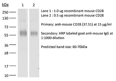
Specifications
| Isotype | Syrian Hamster IgG2 |
|---|---|
| Recommended Isotype Control(s) | InVivoMAb polyclonal Syrian hamster IgG |
| Recommended Dilution Buffer | InVivoPure pH 6.0T Dilution Buffer |
| Conjugation | This product is unconjugated. Conjugation is available via our Antibody Conjugation Services. |
| Immunogen | C57BL/6 mouse T cell lymphoma EL-4 cells |
| Reported Applications |
in vitro T cell stimulation/activation in vivo CD28 blockade |
| Formulation |
PBS, pH 6.0 0.01% Tween Contains no stabilizers or preservatives |
| Endotoxin |
≤1EU/mg (≤0.001EU/μg) Determined by LAL assay |
| Purity |
≥95% Determined by SDS-PAGE |
| Sterility | 0.2 µm filtration |
| Production | Purified from cell culture supernatant in an animal-free facility |
| Purification | Protein G |
| RRID | AB_1107624 |
| Molecular Weight | 150 kDa |
| Storage | The antibody solution should be stored at the stock concentration at 4°C. Do not freeze. |
| Need a Custom Formulation? | See All Antibody Customization Options |
Application References
-
Tang, W., et al (2014). "The oncoprotein and transcriptional regulator Bcl-3 governs plasticity and pathogenicity of autoimmune T cells" Immunity 41(4): 555-566.
PubMed
Bcl-3 is an atypical member of the IkappaB family that modulates transcription in the nucleus via association with p50 (NF-kappaB1) or p52 (NF-kappaB2) homodimers. Despite evidence attesting to the overall physiologic importance of Bcl-3, little is known about its cell-specific functions or mechanisms. Here we demonstrate a T-cell-intrinsic function of Bcl-3 in autoimmunity. Bcl-3-deficient T cells failed to induce disease in T cell transfer-induced colitis and experimental autoimmune encephalomyelitis. The protection against disease correlated with a decrease in Th1 cells that produced the cytokines IFN-gamma and GM-CSF and an increase in Th17 cells. Although differentiation into Th1 cells was not impaired in the absence of Bcl-3, differentiated Th1 cells converted to less-pathogenic Th17-like cells, in part via mechanisms involving expression of the RORgammat transcription factor. Thus, Bcl-3 constrained Th1 cell plasticity and promoted pathogenicity by blocking conversion to Th17-like cells, revealing a unique type of regulation that shapes adaptive immunity.
-
Rabenstein, H., et al (2014). "Differential kinetics of antigen dependency of CD4+ and CD8+ T cells" J Immunol 192(8): 3507-3517.
PubMed
Ag recognition via the TCR is necessary for the expansion of specific T cells that then contribute to adaptive immunity as effector and memory cells. Because CD4+ and CD8+ T cells differ in terms of their priming APCs and MHC ligands we compared their requirements of Ag persistence during their expansion phase side by side. Proliferation and effector differentiation of TCR transgenic and polyclonal mouse T cells were thus analyzed after transient and continuous TCR signals. Following equally strong stimulation, CD4+ T cell proliferation depended on prolonged Ag presence, whereas CD8+ T cells were able to divide and differentiate into effector cells despite discontinued Ag presentation. CD4+ T cell proliferation was neither affected by Th lineage or memory differentiation nor blocked by coinhibitory signals or missing inflammatory stimuli. Continued CD8+ T cell proliferation was truly independent of self-peptide/MHC-derived signals. The subset divergence was also illustrated by surprisingly broad transcriptional differences supporting a stronger propensity of CD8+ T cells to programmed expansion. These T cell data indicate an intrinsic difference between CD4+ and CD8+ T cells regarding the processing of TCR signals for proliferation. We also found that the presentation of a MHC class II-restricted peptide is more efficiently prolonged by dendritic cell activation in vivo than a class I bound one. In summary, our data demonstrate that CD4+ T cells require continuous stimulation for clonal expansion, whereas CD8+ T cells can divide following a much shorter TCR signal.
-
Gu, A. D., et al (2015). "A critical role for transcription factor Smad4 in T cell function that is independent of transforming growth factor beta receptor signaling" Immunity 42(1): 68-79.
PubMed
Transforming growth factor-beta (TGF-beta) suppresses T cell function to maintain self-tolerance and to promote tumor immune evasion. Yet how Smad4, a transcription factor component of TGF-beta signaling, regulates T cell function remains unclear. Here we have demonstrated an essential role for Smad4 in promoting T cell function during autoimmunity and anti-tumor immunity. Smad4 deletion rescued the lethal autoimmunity resulting from transforming growth factor-beta receptor (TGF-betaR) deletion and compromised T-cell-mediated tumor rejection. Although Smad4 was dispensable for T cell generation, homeostasis, and effector function, it was essential for T cell proliferation after activation in vitro and in vivo. The transcription factor Myc was identified to mediate Smad4-controlled T cell proliferation. This study thus reveals a requirement of Smad4 for T-cell-mediated autoimmunity and tumor rejection, which is beyond the current paradigm. It highlights a TGF-betaR-independent role for Smad4 in promoting T cell function, autoimmunity, and anti-tumor immunity.
-
Choi, Y. S., et al (2015). "LEF-1 and TCF-1 orchestrate TFH differentiation by regulating differentiation circuits upstream of the transcriptional repressor Bcl6" Nat Immunol 16(9): 980-990.
PubMed
Follicular helper T cells (TFH cells) are specialized effector CD4(+) T cells that help B cells develop germinal centers (GCs) and memory. However, the transcription factors that regulate the differentiation of TFH cells remain incompletely understood. Here we report that selective loss of Lef1 or Tcf7 (which encode the transcription factor LEF-1 or TCF-1, respectively) resulted in TFH cell defects, while deletion of both Lef1 and Tcf7 severely impaired the differentiation of TFH cells and the formation of GCs. Forced expression of LEF-1 enhanced TFH differentiation. LEF-1 and TCF-1 coordinated such differentiation by two general mechanisms. First, they established the responsiveness of naive CD4(+) T cells to TFH cell signals. Second, they promoted early TFH differentiation via the multipronged approach of sustaining expression of the cytokine receptors IL-6Ralpha and gp130, enhancing expression of the costimulatory receptor ICOS and promoting expression of the transcriptional repressor Bcl6.
Product Citations
-
IL-6 Exacerbates Experimental Autoimmune Prostatitis by Disrupting STAT5a-Mediated Treg Cell Function and Th17/Treg Balance.
In Mediators Inflamm on 26 May 2026 by Liu, X., Bian, X., et al.
PubMed
Chronic prostatitis/chronic pelvic pain syndrome (CP/CPPS) is a common urological condition in men. Although disruption of the Th17 (T helper 17)/Treg (regulatory T) balance has been implicated in its pathogenesis, the upstream drivers of this immune imbalance remain incompletely understood. This study investigated whether interleukin-6 (IL-6) is associated with altered Th17/Treg homeostasis and impaired Treg function in experimental autoimmune prostatitis (EAP).
-
Cardiolipin preserves Treg metabolic fitness and immune homeostasis in the gut.
In Nat Metab on 18 May 2026 by Regina, A., Solagna, F., et al.
PubMed
Loss of host-microbiota balance promotes gut inflammation, colitis and inflammatory bowel disease. Yet, whether host or microbial factors are the critical driver of the pathology remains unclear. Here, we investigate how cardiolipin maintains metabolic fitness of regulatory T (Treg) cells to preserve gut-immune homeostasis. We discover that deleting the cardiolipin-synthesizing enzyme protein tyrosine phosphatase mitochondrial 1 (PTPMT1) in T cells predisposes mice to colitis due to impaired Treg cell function in the absence of dysbiosis. Subsequent pathobiont infections accelerate the progression and severity of gut inflammation. Mechanistically, the absence of cardiolipin impairs Treg cell metabolic fitness and triggers a maladaptive integrated stress response, which can be reversed pharmacologically or genetically, restoring gut homeostasis and extending lifespan in PTPMT1 ΔT mice. Barth syndrome, a genetic disorder marked by severe cardiolipin deficiency, also exhibits gastrointestinal symptoms and inflammation associated with helper T cell imbalance and an active integrated stress response signature. Overall, these results suggest that a cardiolipin-mediated mitonuclear axis in T cells preserves gut-immune homeostasis and dictates outcome in pathobiont infections.
-
Facile induction of immune tolerance by an interleukin-2-TGFβ surrogate agonist.
In Nature on 1 May 2026 by Sun, Q., Barrett, A. K., et al.
PubMed
CD4+ regulatory T cells (Treg cells) are essential for immune tolerance1. Peripherally induced Treg cells (pTreg cells) complement thymic Treg cells by broadening Treg cell reactivity in response to a changing antigenic landscape2. Although both TGFβ and IL-2 synergistically promote functional pTreg cell development in vitro3-6, their combined roles in inducing pTreg cell generation in vivo have not been exploited for tolerizing immunotherapy. Here we designed an IL-2-TGFβ 'surrogate' co-agonist by creating a single-chain fusion protein between IL-2 and a low-affinity TGFβ mimic agonist derived from a helminth parasite7. This IL-2-TGFβ surrogate functions as an AND-gated co-agonist and enabled simultaneous cis-activation of IL-2-STAT5 and TGFβ-SMAD2/3 signalling specifically in T cells that express IL-2 receptors. The IL-2-TGFβ surrogate agonist robustly induced antigen-specific, functional and stable pTreg cells in vivo within peripheral lymphoid organs in mice immunized with ovalbumin (OVA) and myelin oligodendrocyte glycoprotein (MOG)35-55. The induced pTreg cells display an effector-like, actively expanding state with high RORγt expression, enabling efficient migration and suppression of intestinal inflammation. Treatment with this agonist effectively quelled immune activation in mouse models of allergen-induced allergic inflammation and self-antigen-driven autoimmune neuroinflammation, suggesting a strategy for the induction of antigen-specific pTreg cells in vivo to establish immune tolerance in inflammatory, allergic and autoimmune diseases.
-
A senescent tumor cell-derived nanovesicle directly primes splenic T cells to potentiate cancer radiotherapy.
In Cell Rep Med on 21 April 2026 by Pan, J., Zhang, S., et al.
PubMed
Radiotherapy (RT)-induced senescent tumor cells (STCs) reinforce an immunosuppressive tumor microenvironment (ITM) and compromise therapeutic outcomes. However, current senolytic strategies lack specificity for STCs and often cause off-target toxicity. Here, we observe that STCs possess enhanced antigen-presenting capacity in patient-derived tumor tissues and murine tumor models. Leveraging this phenomenon, we engineer STC-derived nanovesicles (termed nano-APM) for preserving endogenous antigens and antigen-presenting cues. We demonstrate that systemically administered nano-APMs accumulate in the spleen and establish a pool of STC-specific CD8+ T cells. Sequential integration of RT induces local tumor senescence, and nano-APMs then effectively mobilize the STC-specific T cells to stimulate a confined recall response. In murine tumor models, the combination of nano-APM plus RT selectively eliminates STCs, reprograms RT-induced ITM, and elicits durable antitumor immunity. Collectively, this study establishes STC-derived nanovesicles as a practical means to enhance RT efficacy by enabling splenic T cell priming and spatiotemporally confined senolysis.