InVivoMAb anti-mouse CD200 (OX2)
Product Description
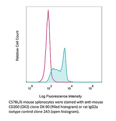
Specifications
| Isotype | Rat IgG2a, κ |
|---|---|
| Recommended Isotype Control(s) | InVivoMAb rat IgG2a isotype control, anti-trinitrophenol |
| Recommended Dilution Buffer | InVivoPure pH 7.0 Dilution Buffer |
| Conjugation | This product is unconjugated. Conjugation is available via our Antibody Conjugation Services. |
| Immunogen | Fusion protein consisting of mouse CD200 (extracellular region) and rat CD4 (domains 3 and 4) |
| Reported Applications |
in vivo CD200 blockade in vitro CD200 blockade Immunohistochemistry (frozen) Immunofluorescence Flow cytometry |
| Formulation |
PBS, pH 7.0 Contains no stabilizers or preservatives |
| Endotoxin |
≤1EU/mg (≤0.001EU/μg) Determined by LAL assay |
| Purity |
≥95% Determined by SDS-PAGE |
| Sterility | 0.2 µm filtration |
| Production | Purified from cell culture supernatant in an animal-free facility |
| Purification | Protein G |
| RRID | AB_2687821 |
| Molecular Weight | 150 kDa |
| Storage | The antibody solution should be stored at the stock concentration at 4°C. Do not freeze. |
| Need a Custom Formulation? | See All Antibody Customization Options |
Application References
-
Choueiry, F., et al (2020). "CD200 promotes immunosuppression in the pancreatic tumor microenvironment" J Immunother Cancer 8(1).
PubMed
BACKGROUND: A significant challenge to overcome in pancreatic ductal adenocarcinoma (PDAC) is the profound systemic immunosuppression that renders this disease non-responsive to immunotherapy. Our supporting data provide evidence that CD200, a regulator of myeloid cell activity, is expressed in the PDAC microenvironment. Additionally, myeloid-derived suppressor cells (MDSC) isolated from patients with PDAC express elevated levels of the CD200 receptor (CD200R). Thus, we hypothesize that CD200 expression in the PDAC microenvironment limits responses to immunotherapy by promoting expansion and activity of MDSC. METHODS: Immunofluorescent staining was used to determine expression of CD200 in murine and human PDAC tissue. Flow cytometry was utilized to test for CD200R expression by immune populations in patient blood samples. In vivo antibody blocking of CD200 was conducted in subcutaneous MT-5 tumor-bearing mice and in a genetically engineered PDAC model (KPC-Brca2 mice). Peripheral blood mononuclear cells (PBMC) from patients with PDAC were analyzed by single-cell RNA sequencing. MDSC expansion assays were completed using healthy donor PBMC stimulated with IL-6/GM-CSF in the presence of recombinant CD200 protein. RESULTS: We found expression of CD200 by human pancreatic cell lines (BxPC3, MiaPaca2, and PANC-1) as well as on primary epithelial pancreatic tumor cells and smooth muscle actin+ stromal cells. CD200R expression was found to be elevated on CD11b+CD33+HLA-DR(lo/-) MDSC immune populations from patients with PDAC (p=0.0106). Higher expression levels of CD200R were observed in CD15+ MDSC compared with CD14+ MDSC (p<0.001). In vivo studies demonstrated that CD200 antibody blockade limited tumor progression in MT-5 subcutaneous tumor-bearing and in KPC-Brca2 mice (p<0.05). The percentage of intratumoral MDSC was significantly reduced in anti-CD200 treated mice compared with controls. Additionally, in vivo blockade of CD200 can also significantly enhance the efficacy of PD-1 checkpoint antibodies compared with single antibody therapies (p<0.05). Single-cell RNA sequencing of PBMC from patients revealed that CD200R+ MDSC expressed genes involved in cytokine signaling and MDSC expansion. Further, in vitro cytokine-driven expansion and the suppressive activity of human MDSC was enhanced when cocultured with recombinant CD200 protein. CONCLUSIONS: These results indicate that CD200 expression in the PDAC microenvironment may regulate MDSC expansion and that targeting CD200 may enhance activity of checkpoint immunotherapy.
-
Seeds, R. E., et al (2011). "The role of myeloid receptors on murine plasmacytoid dendritic cells in induction of type I interferon" Int Immunopharmacol 11(7): 794-801.
PubMed
This study tested the hypothesis that a set of predominantly myeloid restricted receptors (F4/80, CD36, Dectin-1, CD200 receptor and mannan binding lectins) and the broadly expressed CD200 played a role in a key function of plasmacytoid DC (pDC), virally induced type I interferon (IFN) production. The Dectin-1 ligands zymosan, glucan phosphate and the anti-Dectin-1 monoclonal antibody (mAb) 2A11 had no effect on influenza virus induced IFNalpha/beta production by murine splenic pDC. However, mannan, a broad blocking reagent against mannose specific receptors, inhibited IFNalpha/beta production by pDC in response to inactivated influenza virus. Moreover, viral glycoproteins (influenza virus haemagglutinin and HIV-1 gp120) stimulated IFNalpha/beta production by splenocytes in a mannan-inhibitable manner, implicating the function of a lectin in glycoprotein induced IFN production. Lastly, the effect of CD200 on IFN induction was investigated. CD200 knock-out macrophages produced more IFNalpha than wild-type macrophages in response to polyI:C, a MyD88-independent stimulus, consistent with CD200’s known inhibitory effect on myeloid cells. In contrast, blocking CD200 with an anti-CD200 mAb resulted in reduced IFNalpha production by pDC-containing splenocytes in response to CpG and influenza virus (MyD88-dependent stimuli). This suggests there could be a differential effect of CD200 on MyD88 dependent and independent IFN induction pathways in pDC and macrophages. This study supports the hypothesis that a mannan-inhibitable lectin and CD200 are involved in virally induced type I IFN induction.
-
Koning, N., et al (2009). "Distribution of the immune inhibitory molecules CD200 and CD200R in the normal central nervous system and multiple sclerosis lesions suggests neuron-glia and glia-glia interactions" J Neuropathol Exp Neurol 68(2): 159-167.
PubMed
CD200 is a membrane glycoprotein that suppresses immune activity via its receptor, CD200R. CD200-CD200R interactions have recently been considered to contribute to the “immune privileged” status of the central nervous system (CNS). The mechanisms by which these interactions take place are not well understood in part because there is limited detailed information on the distribution of CD200 and CD200R in the CNS. Here, we used immunohistochemistry to characterize the distinct anatomical and cellular distribution of these molecules in multiple sclerosis (MS) lesions and controls. CD200 was robustly expressed in gray matter areas including the cerebral cortex, hippocampus, striatum, cerebellum, and spinal cord, where neurons appeared immunopositive. CD200 expression was also detected in oligodendrocytes, but not in astrocytes or microglia. In CNS samples from MS patients, CD200 expression was additionally observed on reactive astrocytes in chronic active plaque centers, despite our previous finding of an overall decrease ofCD200 expression in MS lesions. In contrast to CD200, the immunolocalization pattern of CD200R was very distinct, showing high expression on perivascular macrophages in both gray and white matter. Using flow cytometry, we also found that human primary microglia express low levels of CD200R. These data suggest that CD200-mediated immune suppression may occur not only via neuron-microglia interactions, but also via glia-glia interactions, especially in inflammatory conditions in which an immune-suppressive environment needs to be restored; this may occur as a result of increased CD200 expression on reactive astrocytes.
-
Snelgrove, R. J., et al (2008). "A critical function for CD200 in lung immune homeostasis and the severity of influenza infection" Nat Immunol 9(9): 1074-1083.
PubMed
The lung must maintain a high threshold of immune ‘ignorance’ to innocuous antigens to avoid inflammatory disease that depends on the balance of positive inflammatory signals and repressor pathways. We demonstrate here that airway macrophages had higher expression of the negative regulator CD200 receptor (CD200R) than did their systemic counterparts. Lung macrophages were restrained by CD200 expressed on airway epithelium. Mice lacking CD200 had more macrophage activity and enhanced sensitivity to influenza infection, which led to delayed resolution of inflammation and, ultimately, death. The administration of agonists that bind CD200R, however, prevented inflammatory lung disease. Thus, CD200R is critical for lung macrophage immune homeostasis in the resting state and limits inflammatory amplitude and duration during pulmonary influenza infection.
Product Citations
-
Single-Cell Transcriptional Heterogeneity of Lymphatic Endothelial Cells in Normal and Inflamed Murine Lymph Nodes.
In Cells on 2 June 2021 by Sibler, E., He, Y., et al.
PubMed
The lymphatic system plays a crucial role in immunity and lymph nodes (LNs) undergo drastic remodeling during inflammation. Here, we used single-cell RNA sequencing to investigate transcriptional changes in lymphatic endothelial cells (LECs) in LNs draining naïve and inflamed skin. We found that subsets of LECs lining the different LN sinuses responded individually to skin inflammation, suggesting that they exert distinct functions under pathological conditions. Among the genes dysregulated during inflammation, we confirmed an up-regulation of CD200 in the LECs lining the subcapsular sinus floor with a possible function in immune regulation. Furthermore, by in silico analysis, we predicted numerous possible interactions of LECs with diverse immune cells in the LNs and found similarities in the transcriptional changes of LN LECs in different skin inflammation settings. In summary, we provide an in-depth analysis of the transcriptional landscape of LN LECs in the naïve state and in skin inflammation.