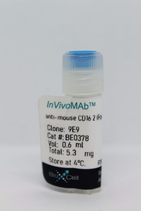
InVivoMAb anti-mouse CD16.2 (FcγRIV)
Product Details
The 9E9 monoclonal antibody reacts with mouse CD16.2, also known as FcγRIV (Fc receptor, IgG, low affinity IV). CD16.2 is a member of the immunoglobulin superfamily and is expressed on monocytes, macrophages, dendritic cells, and neutrophils. Fcγ receptors are essential for IgG-dependent effector functions in vivo. CD16.2 requires the common Fcγ chain for expression and signaling. CD16.2 binds to IgG2a and IgG2b with intermediate affinity. IgG2a- and IgG2b-dependent effector functions are severely impaired in CD16.2 deficient mice. CD16.2 has also been reported to be a low-affinity IgE receptor for all IgE allotypes and promotes IgE-induced lung inflammation. The 9E9 antibody has been shown to inhibit cellular CD16.2 function.Specifications
| Isotype | Armenian hamster IgG |
|---|---|
| Recommended Isotype Control(s) | InVivoMAb polyclonal Armenian hamster IgG |
| Recommended Dilution Buffer | InVivoPure pH 7.0 Dilution Buffer |
| Conjugation | This product is unconjugated. Conjugation is available via our Antibody Conjugation Services. |
| Immunogen | Mouse FcγRIV extracellular domain-mouse IgG1 Fc fusion protein |
| Reported Applications |
in vivo CD16.2 blockade in vitro CD16.2 blockade Flow cytometry |
| Formulation |
PBS, pH 7.0 Contains no stabilizers or preservatives |
| Endotoxin |
<2EU/mg (<0.002EU/μg) Determined by LAL gel clotting assay |
| Purity |
>95% Determined by SDS-PAGE |
| Sterility | 0.2 μM filtered |
| Purification | Protein A |
| RRID | AB_2927515 |
| Molecular Weight | 150 kDa |
| Storage | The antibody solution should be stored at the stock concentration at 4°C. Do not freeze. |
Recommended Products
-
Recommended Isotype Control(s)
InVivoMAb polyclonal Armenian hamster IgG
-

Recommended Dilution Buffer
InVivoPure pH 7.0 Dilution Buffer
in vivo CD16.2 blockade
Gordan, S., et al. (2019). "The Immunological Organ Environment Dictates the Molecular and Cellular Pathways of Cytotoxic Antibody Activity" Cell Rep 29(10): 3033-3046 e3034. PubMed
Cytotoxic immunoglobulin G antibodies are an essential component of therapeutic approaches aimed at depleting self-reactive or malignant cells. More recent evidence suggests that the tissue in which the target cell resides influences the underlying molecular and cellular pathways responsible for cytotoxic antibody activity. By studying cytotoxic IgG activity directed against natural killer cells in primary and secondary immunological organs, we show that distinct organ-specific effector pathways are responsible for target cell depletion. While in the bone marrow, the classical complement pathway and the high-affinity Fcgamma-receptor I expressed on organ-resident macrophages were both involved in removing opsonized target cells; in the spleen and blood, all activating FcgammaRs but not the classical complement pathway were critical for target cell killing. Our study suggests that future strategies aimed at optimizing overall cytotoxic antibody activity may need to consider organ-specific pathways to achieve a maximal therapeutic effect.
in vivo CD16.2 blockade
Schulze, F. S., et al. (2014). "Fcgamma receptors III and IV mediate tissue destruction in a novel adult mouse model of bullous pemphigoid" Am J Pathol 184(8): 2185-2196. PubMed
Bullous pemphigoid (BP) and epidermolysis bullosa acquisita are subepidermal autoimmune blistering diseases mediated by autoantibodies against type XVII collagen (Col17) and Col7, respectively. For blister formation, Fc-mediated events, such as infiltration of inflammatory cells in the skin, complement activation, and release of proteases at the dermal-epidermal junction, are essential. Although in the neonatal passive transfer mouse model of BP, tissue destruction is mediated by Fcgamma receptors (FcgammaRs) I and III, the passive transfer model of epidermolysis bullosa acquisita completely depends on FcgammaRIV. To clarify this discrepancy, we developed a novel experimental model for BP using adult mice. Lesion formation was Fc mediated because gamma-chain-deficient mice and mice treated with anti-Col17 IgG, depleted from its sugar moiety at the Fc portion, were resistant to disease induction. By the use of various FcgammaR-deficient mouse strains, tissue destruction was shown to be mediated by FcgammaRIV, FcgammaRIII, and FcgammaRIIB, whereas FcgammaRI was not essential. Furthermore, anti-inflammatory mediators in already clinically diseased mice can be explored in the novel BP model, because the pharmacological inhibition of FcgammaRIV and depletion of granulocytes abolished skin blisters. Herein, we extended our knowledge about the importance of FcgammaRs in experimental BP and established a novel BP mouse model suitable to study disease development over a longer time period and explore novel treatment strategies in a quasi-therapeutic setting.
in vivo CD16.2 blockade, in vitro CD16.2 blockade, Flow Cytometry
Syed, S. N., et al. (2009). "Both FcgammaRIV and FcgammaRIII are essential receptors mediating type II and type III autoimmune responses via FcRgamma-LAT-dependent generation of C5a" Eur J Immunol 39(12): 3343-3356. PubMed
FcgammaRIV is a relatively new IgG Fc receptor (FcgammaR) that is reported to contribute to the pathogenesis of autoimmune diseases, although its specific role in relation to FcgammaRIII, complement and IgG2 subclasses remains uncertain. Here we define FcgammaRIV on macrophages as a receptor for soluble IgG2a/b complexes but not for cellular bound IgG2a and show that simultaneous activation of FcgammaRIV and FcgammaRIII is critical to mediate certain type II/III autoimmune responses. FcgammaRIII-deficient mice display compensatory enhanced FcgammaRIV expression, are protected from lung inflammation after deposition of IgG complexes, and show reduced sensitivity to IgG2a/b-mediated hemolytic anemia, indicating that increased FcgammaRIV alone is not sufficient to trigger these diseases in the absence of FcgammaRIII. Importantly, however, blockade of FcgammaRIV is also effective in inhibiting phagocytosis and cytokine production in IgG2b-induced anemia and acute lung injury, processes that display a further dependence on C5a anaphylatoxin receptor. Using gene deletion and functional inhibition studies, we found that FcgammaRIII and FcgammaRIV are each essential to trigger an FcRgamma-linker for activation of T-cell-dependent signal that drives C5a production in the Arthus reaction. Together, the results demonstrate a combined requirement for FcgammaRIII and FcgammaRIV in autoimmune injury, and identify the linker for activation of T cells adaptor as an integral component of linked FcgammaR and C5a anaphylatoxin receptor activation to generate inflammation.
in vivo CD16.2 blockade, in vitro CD16.2 blockade, Flow Cytometry
Mancardi, D. A., et al. (2008). "FcgammaRIV is a mouse IgE receptor that resembles macrophage FcepsilonRI in humans and promotes IgE-induced lung inflammation" J Clin Invest 118(11): 3738-3750. PubMed
FcgammaRIV is a recently identified mouse activating receptor for IgG2a and IgG2b that is expressed on monocytes, macrophages, and neutrophils; herein it is referred to as mFcgammaRIV. Although little is known about mFcgammaRIV, it has been proposed to be the mouse homolog of human FcgammaRIIIA (hFcgammaRIIIA) because of high sequence homology. Our work, however, has revealed what we believe to be new properties of mFcgammaRIV that endow this receptor with a previously unsuspected biological significance; we have shown that it is a low-affinity IgE receptor for all IgE allotypes. Although mFcgammaRIV functioned as a high-affinity IgG receptor, mFcgammaRIV-bound monomeric IgGs were readily displaced by IgE immune complexes. Engagement of mFcgammaRIV by IgE immune complexes induced bronchoalveolar and peritoneal macrophages to secrete cytokines, suggesting that mFcgammaRIV may be an equivalent of human FceRI(alphagamma), which is expressed by macrophages and neutrophils and especially in atopic individuals, rather than an equivalent of hFcgammaRIIIA, which has no affinity for IgE. Using mice lacking 3 FcgammaRs and 2 FceRs and expressing mFcgammaRIV only, we further demonstrated that mFcgammaRIV promotes IgE-induced lung inflammation. These data lead us to propose a mouse model of IgE-induced lung inflammation in which cooperation exists between mast cells and mFcgammaRIV-expressing lung cells. We therefore suggest that a similar cooperation may occur between mast cells and hFceRI-expressing lung cells in human allergic asthma.