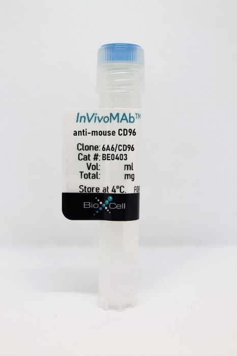
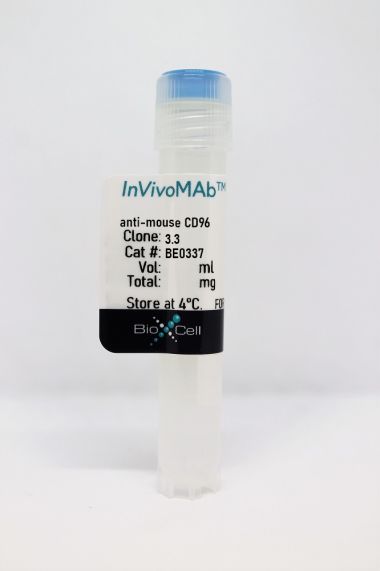
InVivoMAb anti-mouse CD96
Product Details
The 6A6 monoclonal antibody reacts with mouse CD96, also known as T cell activation increased late expression (TACTILE) in humans. CD96 is a type I transmembrane glycoprotein immunoglobulin (Ig) superfamily receptor with a complex extracellular domain having three Ig-like domains and one cytoplasmic domain. CD96 is expressed at low levels on resting NK cells and T cells and at high levels on activated NK and T cells. CD96 binds to its high-affinity ligand, the CD155/poliovirus receptor (PVR), thereby triggering T cell and NK cell adhesion and function. CD96 is also involved in the negative regulation of NK cell-mediated immune surveillance. CD96 has recently been identified as a novel target for cancer immunotherapy and has been shown to play a role in metastasis. In vitro experiments with CD96-blocking antibodies (clones 6A6 and 3.3) and a non-blocking antibody (clone 8B10) have established that the 6A6 antibody binds to the first Ig domain of murine CD96 with superior affinity and that the 6A6 antibody competes with CD155 binding, thereby blocking the CD96-CD155 interaction. In a mouse model of pancreatic ductal adenocarcinoma (PDAC), in vivo blockade of CD96 with 6A6 after neutralization of PD-1 with the RMP1-14 antibody led to the prevention of post-surgery PDAC recurrence while facilitating long-term survival. Mechanistic experiments with in vivo use of 6A6 and other CD96 antibodies on CD155-deficient mice and CD226-deficient mice showed that the CD96 antibodies need not block CD96-CD155 interactions to promote NK cell anti-metastatic activity in tumors.Specifications
| Isotype | Rat IgG2a, λ |
|---|---|
| Recommended Isotype Control(s) | InVivoMAb rat IgG2a isotype control, anti-trinitrophenol |
| Recommended Dilution Buffer | InVivoPure pH 7.0 Dilution Buffer |
| Conjugation | This product is unconjugated. Conjugation is available via our Antibody Conjugation Services. |
| Immunogen | mCD96-hIgG fusion protein |
| Reported Applications |
in vivo blocking of CD96 in vitro blocking of CD96 Functional assays Flow cytometry |
| Formulation |
PBS, pH 7.0 Contains no stabilizers or preservatives |
| Endotoxin |
<2EU/mg (<0.002EU/μg) Determined by LAL gel clotting assay |
| Purity |
>95% Determined by SDS-PAGE |
| Sterility | 0.2 µm filtration |
| Production | Purified from cell culture supernatant in an animal-free facility |
| Purification | Protein G |
| Molecular Weight | 150 kDa |
| Storage | The antibody solution should be stored at the stock concentration at 4°C. Do not freeze. |
Recommended Products
in vitro blocking of CD96
Luo B, Sun Y, Zhan Q, Luo Y, Chen Y, Fu T, Yang T, Ren L, Xie Z, Situ X, Liu B, Tang K, Ke Z. (2024). "Combining TIGIT blockade with IL-15 stimulation is a promising immunotherapy strategy for lung adenocarcinoma" Clin Transl Med 14(1):e1553. PubMed
Background: T-cell immunoglobulin and immunoreceptor tyrosine-based inhibitory motif domain (TIGIT) is an immune checkpoint molecule that suppresses CD8+ T-cell function in cancer. However, the expression profile and functional significance of TIGIT in the immune microenvironment of lung adenocarcinoma (LUAD) remain elusive. Interleukin (IL)-15 has emerged as a promising candidate for enhancing CD8+ T-cell mediated tumour eradication. Exploring therapeutic strategies that combine IL-15 with TIGIT blockade in LUAD is warranted. Methods: We investigated the regulatory network involving coinhibitory TIGIT and CD96, as well as costimulatory CD226 in LUAD using clinical samples. The potential role of TIGIT in regulating the pathogenesis of LUAD was addressed through a murine model with transplanted tumours constructed in Tigit-/- mice. The therapeutic strategy that combines TIGIT blockade with IL-15 stimulation was verified using a transplanted tumour murine model and a patient-derived organoid (PDO) model. Results: The frequency of TIGIT+ CD8+ T cells was significantly increased in LUAD. Increased TIGIT expression indicated poorer prognosis in LUAD patients. Furthermore, the effector function of TIGIT+ CD8+ tumour-infiltrating lymphocytes (TILs) was impaired in LUAD patients and TIGIT inhibited antitumour immune response of CD8+ TILs in tumour-bearing mice. Mechanistically, IL-15 enhanced the effector function of CD8+ TILs but stimulated the expression of TIGIT on CD8+ TILs concomitantly. The application of IL-15 combined with TIGIT blockade showed additive effects in enhancing the cytotoxicity of CD8+ TILs and thus further increased the antitumour immune response in LUAD. Conclusions: Our findings identified TIGIT as a promising therapeutic target for LUAD. LUAD could benefit more from the combined therapy of IL-15 stimulation and TIGIT blockade.
in vitro CD96 blocking
Chiang EY, de Almeida PE, de Almeida Nagata DE, Bowles KH, Du X, Chitre AS, Banta KL, Kwon Y, McKenzie B, Mittman S, Cubas R, Anderson KR, Warming S, Grogan JL. (2020). "CD96 functions as a co-stimulatory receptor to enhance CD8+ T cell activation and effector responses" Eur J Immunol 50(6):891-902. PubMed
CD96 is a member of the poliovirus receptor (PVR, CD155)-nectin family that includes T cell Ig and ITIM domain (TIGIT) and CD226. While CD96, TIGIT, and CD226 have important roles in regulating NK cell activity, and TIGIT and CD226 have also been shown to regulate T cell responses, it is unclear whether CD96 has inhibitory or stimulatory function in CD8+ T cells. Here, we demonstrate that CD96 has co-stimulatory function on CD8+ T cells. Crosslinking of CD96 on human or mouse CD8+ T cells induced activation, effector cytokine production, and proliferation. CD96 was found to transduce its activating signal through the MEK-ERK pathway. CD96-mediated signaling led to increased frequencies of NUR77- and T-bet-expressing CD8+ T cells and enhanced cytotoxic effector activity, indicating that CD96 can modulate effector T cell differentiation. Antibody blockade of CD96 or genetic ablation of CD96 expression on CD8+ T cells impaired expression of transcription factors and proinflammatory cytokines associated with CD8+ T cell activation in in vivo models. Taken together, CD96 has a co-stimulatory role in CD8+ T cell activation and effector function.
in vitro functional assay
Okumura G, Iguchi-Manaka A, Murata R, Yamashita-Kanemaru Y, Shibuya A, Shibuya K. (2020). "Tumor-derived soluble CD155 inhibits DNAM-1-mediated antitumor activity of natural killer cells" J Exp Med 217(4):1. PubMed
CD155 is a ligand for DNAM-1, TIGIT, and CD96 and is involved in tumor immune responses. Unlike mouse cells, human cells express both membranous CD155 and soluble CD155 (sCD155) encoded by splicing isoforms of CD155. However, the role of sCD155 in tumor immunity remains unclear. Here, we show that, after intravenous injection with sCD155-producing B16/BL6 melanoma, the numbers of tumor colonies in wild-type (WT), TIGIT knock-out (KO), or CD96 KO mice, but not DNAM-1 KO mice, were greater than after injection with parental B16/BL6 melanoma. NK cell depletion canceled the difference in the numbers of tumor colonies in WT mice. In vitro assays showed that sCD155 interfered with DNAM-1-mediated NK cell degranulation. In addition, DNAM-1 had greater affinity than TIGIT and CD96 for sCD155, suggesting that sCD155 bound preferentially to DNAM-1. Together, these results demonstrate that sCD155 inhibits DNAM-1-mediated cytotoxic activity of NK cells, thus promoting the lung colonization of B16/BL6 melanoma.
in vivo blocking of CD96, in vitro functional assay, Flow Cytometry
Roman Aguilera A, Lutzky VP, Mittal D, Li XY, Stannard K, Takeda K, Bernhardt G, Teng MWL, Dougall WC, Smyth MJ. (2018). "CD96 targeted antibodies need not block CD96-CD155 interactions to promote NK cell anti-metastatic activity" Oncoimmunology 7(5):e1424677. PubMed
CD96 is a transmembrane glycoprotein Ig superfamily receptor, expressed on various T cell subsets and NK cells, that interacts with nectin and nectin-like proteins, including CD155/polio virus receptor (PVR). Here, we have compared three rat anti-mouse CD96 mAbs, including two that block CD96-CD155 (3.3 and 6A6) and one that does not block CD96-CD155 (8B10). Using flow cytometry, we demonstrated that both mAbs 3.3 and 6A6 bind to the first Ig domain of mouse CD96 and compete with CD155 binding, while mAb 8B10 binds to the second Ig domain and does not block CD155. While Fc isotype was irrelevant concerning the anti-metastatic activity of 3.3 mAb, in four different experimental metastases models and one spontaneous metastasis model, the relative order of anti-metastatic potency was 6A6 > 3.3 > 8B10. The metastatic burden control of all of the anti-CD96 clones was highly dependent on NK cells and IFN-γ. Consistent with its inability to block CD96-CD155 interactions, 8B10 retained anti-metastatic activity in CD155-deficient mice, whereas 3.3 and 6A6 lost potency in CD155-deficient mice. Furthermore, 8B10 retained most of its anti-metastatic activity in IL-12p35-deficient mice whereas the activity of 3.3 and 6A6 were partially lost. All three mAbs were inactive in CD226-deficient mice. Altogether, these data demonstrate anti-CD96 need not block CD96-CD155 interactions (ie. immune checkpoint blockade) to promote NK cell anti-metastatic activity.
in vivo blocking of CD96
Brooks J, Fleischmann-Mundt B, Woller N, Niemann J, Ribback S, Peters K, Demir IE, Armbrecht N, Ceyhan GO, Manns MP, Wirth TC, Kubicka S, Bernhardt G, Smyth MJ, Calvisi DF, Gürlevik E, Kühnel F. (2018). "Perioperative, Spatiotemporally Coordinated Activation of T and NK Cells Prevents Recurrence of Pancreatic Cancer" Cancer Res 78(2):475-488. PubMed
Pancreatic ductal adenocarcinoma (PDAC) is a highly lethal and disseminating cancer resistant to therapy, including checkpoint immunotherapies, and early tumor resection and (neo)adjuvant chemotherapy fails to improve a poor prognosis. In a transgenic mouse model of resectable PDAC, we investigated the coordinated activation of T and natural killier (NK) cells in addition to gemcitabine chemotherapy to prevent tumor recurrence. Only neoadjuvant, but not adjuvant treatment with a PD-1 antagonist effectively supported chemotherapy and suppressed local tumor recurrence and improved survival involving both NK and T cells. Local T-cell activation was confirmed by increased tumor infiltration with CD103+CD8+ T cells and neoantigen-specific CD8 T lymphocytes against the marker neoepitope LAMA4-G1254V. To achieve effective prevention of distant metastases in a complementary approach, we blocked the NK-cell checkpoint CD96, an inhibitory NK-cell receptor that binds CD155, which was abundantly expressed in primary PDAC and metastases of human patients. In gemcitabine-treated mice, neoadjuvant PD-1 blockade followed by adjuvant inhibition of CD96 significantly prevented relapse of PDAC, allowing for long-term survival. In summary, our results show in an aggressively growing transgenic mouse model of PDAC that the coordinated activation of both innate and adaptive immunity can effectively reduce the risk of tumor recurrence after surgery, facilitating long-term remission of this lethal disease.Significance: Coordinated neoadjuvant and adjuvant immunotherapies reduce the risk of disease relapse after resection of murine PDAC, suggesting this concept for future clinical trials. Cancer Res; 78(2); 475-88. ©2017 AACR.
in vivo blocking of CD96
Blake SJ, Stannard K, Liu J, Allen S, Yong MC, Mittal D, Aguilera AR, Miles JJ, Lutzky VP, de Andrade LF, Martinet L, Colonna M, Takeda K, Kühnel F, Gurlevik E, Bernhardt G, Teng MW, Smyth MJ. (2016). "Suppression of Metastases Using a New Lymphocyte Checkpoint Target for Cancer Immunotherapy" Cancer Discov 6(4):446-59. PubMed
CD96 has recently been shown as a negative regulator of mouse natural killer (NK)-cell activity, with Cd96(-/-)mice displaying hyperresponsive NK cells upon immune challenge. In this study, we have demonstrated that blocking CD96 with a monoclonal antibody inhibited experimental metastases in three different tumor models. The antimetastatic activity of anti-CD96 was dependent on NK cells, CD226 (DNAM-1), and IFNγ, but independent of activating Fc receptors. Anti-CD96 was more effective in combination with anti-CTLA-4, anti-PD-1, or doxorubicin chemotherapy. Blocking CD96 in Tigit(-/-)mice significantly reduced experimental and spontaneous metastases compared with its activity in wild-type mice. Co-blockade of CD96 and PD-1 potently inhibited lung metastases, with the combination increasing local NK-cell IFNγ production and infiltration. Overall, these data demonstrate that blocking CD96 is a new and complementary immunotherapeutic strategy to reduce tumor metastases. Significance: This article illustrates the antimetastatic activity and mechanism of action of an anti-CD96 antibody that inhibits the CD96-CD155 interaction and stimulates NK-cell function. Targeting host CD96 is shown to complement surgery and conventional immune checkpoint blockade.
in vitro CD96 blocking
Stanietsky N, Rovis TL, Glasner A, Seidel E, Tsukerman P, Yamin R, Enk J, Jonjic S, Mandelboim O. (2013). "Mouse TIGIT inhibits NK-cell cytotoxicity upon interaction with PVR" Eur J Immunol 43(8):2138-50. PubMed
The activity of natural killer (NK) cells is controlled by a balance of signals derived from inhibitory and activating receptors. TIGIT is a novel inhibitory receptor, recently shown in humans to interact with two ligands: PVR and Nectin2 and to inhibit human NK-cell cytotoxicity. Whether mouse TIGIT (mTIGIT) inhibits mouse NK-cell cytotoxicity is unknown. Here we show that mTIGIT is expressed by mouse NK cells and interacts with mouse PVR. Using mouse and human Ig fusion proteins we show that while the human TIGIT (hTIGIT) cross-reacts with mouse PVR (mPVR), the binding of mTIGIT is restricted to mPVR. We further demonstrate using surface plasmon resonance (SPR) and staining with Ig fusion proteins that mTIGIT binds to mPVR with higher affinity than the co-stimulatory PVR-binding receptor mouse DNAM1 (mDNAM1). Functionally, we show that triggering of mTIGIT leads to the inhibition of NK-cell cytotoxicity, that IFN-γ secretion is enhanced when mTIGIT is blocked and that the TIGIT-mediated inhibition is dominant over the signals delivered by the PVR-binding co-stimulatory receptors. Additionally, we identify the inhibitory motif responsible for mTIGIT inhibition. In conclusion, we show that TIGIT is a powerful inhibitory receptor for mouse NK cells.
in vitro CD96 blocking, Flow Cytometry
Seth S, Maier MK, Qiu Q, Ravens I, Kremmer E, Förster R, Bernhardt G. (2007). "The murine pan T cell marker CD96 is an adhesion receptor for CD155 and nectin-1" The CD155 ligand CD96 is an immunoglobulin-like protein tentatively allocated to the repertoire of human NK receptors. We report here that the CD96/CD155-interaction is preserved between man and mouse although both receptors are only moderately conserved in amino acid sequence. Moreover, murine CD96 (mCD96) binds to nectin-1, a receptor related to CD155. Applying newly generated monoclonal antibodies specifically recognizing mCD96, an expression profile is revealed resembling closely that of human CD96 (hCD96) on cells of hematopoietic origin. A panel of anti-mCD96 but also recently established anti-mCD155 antibodies effectively prevents formation of CD96/CD155-complexes. This was exploited to demonstrate that the only available receptor for mCD96 present on thymocytes is mCD155. Moreover, T cell adhesion to insect cells expressing mCD155 is blocked by these antibodies depending on the T cell subtype. These results suggest a function of the CD96/CD155-adhesion system in T cell biology. 364(4):959-65. PubMed
The CD155 ligand CD96 is an immunoglobulin-like protein tentatively allocated to the repertoire of human NK receptors. We report here that the CD96/CD155-interaction is preserved between man and mouse although both receptors are only moderately conserved in amino acid sequence. Moreover, murine CD96 (mCD96) binds to nectin-1, a receptor related to CD155. Applying newly generated monoclonal antibodies specifically recognizing mCD96, an expression profile is revealed resembling closely that of human CD96 (hCD96) on cells of hematopoietic origin. A panel of anti-mCD96 but also recently established anti-mCD155 antibodies effectively prevents formation of CD96/CD155-complexes. This was exploited to demonstrate that the only available receptor for mCD96 present on thymocytes is mCD155. Moreover, T cell adhesion to insect cells expressing mCD155 is blocked by these antibodies depending on the T cell subtype. These results suggest a function of the CD96/CD155-adhesion system in T cell biology.


