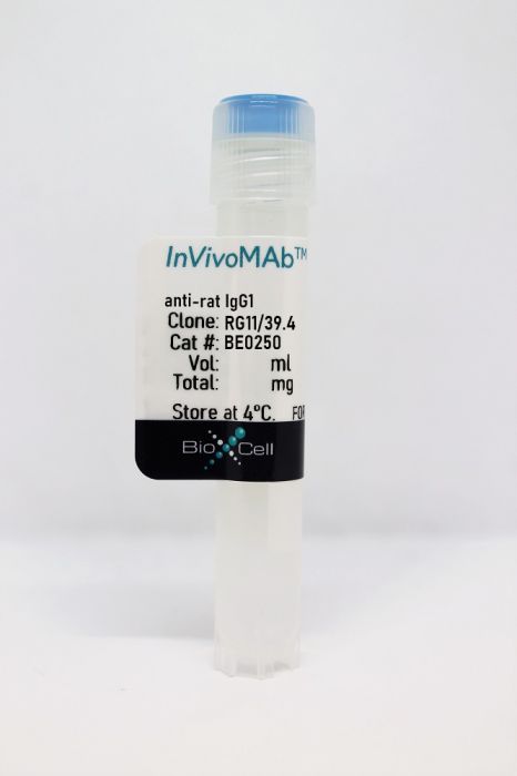
InVivoMAb anti-rat IgG1
Product Details
The RG11/39.4 monoclonal antibody reacts with the Fc region of rat IgG1. It is commonly used as a secondary antibody to detect rat IgG1 antibodies in various diagnostic applications.Specifications
| Isotype | Mouse IgG2c, κ |
|---|---|
| Recommended Isotype Control(s) | InVivoMAb mouse IgG2c isotype control, anti-dengue virus |
| Recommended Dilution Buffer | InVivoPure pH 7.0 Dilution Buffer |
| Conjugation | This product is unconjugated. Conjugation is available via our Antibody Conjugation Services. |
| Immunogen | Rat IgG1 |
| Reported Applications | Flow cytometry |
| Formulation |
PBS, pH 7.0 Contains no stabilizers or preservatives |
| Endotoxin |
<2EU/mg (<0.002EU/μg) Determined by LAL gel clotting assay |
| Purity |
>95% Determined by SDS-PAGE |
| Sterility | 0.2 µm filtration |
| Production | Purified from cell culture supernatant in an animal-free facility |
| Purification | Protein A |
| RRID | AB_2687731 |
| Molecular Weight | 150 kDa |
| Storage | The antibody solution should be stored at the stock concentration at 4°C. Do not freeze. |
Recommended Products
Flow Cytometry
Behler, F., et al. (2015). "Macrophage-inducible C-type lectin Mincle-expressing dendritic cells contribute to control of splenic Mycobacterium bovis BCG infection in mice" Infect Immun 83(1): 184-196. PubMed
The macrophage-inducible C-type lectin Mincle has recently been identified to be a pattern recognition receptor sensing mycobacterial infection via recognition of the mycobacterial cell wall component trehalose-6′,6-dimycolate (TDM). However, its role in systemic mycobacterial infections has not been examined so far. Mincle-knockout (KO) mice were infected intravenously with Mycobacterium bovis BCG to mimic the systemic spread of mycobacteria under defined experimental conditions. After intravenous infection with M. bovis BCG, Mincle-KO mice responded with significantly higher numbers of mycobacterial CFU in spleen and liver, while reduced granuloma formation was observed only in the spleen. At the same time, reduced Th1 cytokine production and decreased numbers of gamma interferon-producing T cells were observed in the spleens of Mincle-KO mice relative to the numbers in the spleens of wild-type (WT) mice. The effect of adoptive transfer of defined WT leukocyte subsets generated from bone marrow cells of zDC(+/DTR) mice (which bear the human diphtheria toxin receptor under the control of the classical dendritic cell-specific zinc finger transcription factor zDC) to specifically deplete Mincle-expressing classical dendritic cells (cDCs) but not macrophages after diphtheria toxin application on the numbers of splenic and hepatic CFU and T cell subsets was then determined. Adoptive transfer experiments revealed that Mincle-expressing splenic cDCs rather than Mincle-expressing macrophages contributed to the reconstitution of attenuated splenic antimycobacterial immune responses in Mincle-KO mice after intravenous challenge with BCG. Collectively, we show that expression of Mincle, particularly by cDCs, contributes to the control of splenic M. bovis BCG infection in mice.
Flow Cytometry
Huang, G., et al. (2014). "Characterization of transfusion-elicited acute antibody-mediated rejection in a rat model of kidney transplantation" Am J Transplant 14(5): 1061-1072. PubMed
Animal models of antibody-mediated rejection (ABMR) may provide important evidence supporting proof of concept. We elicited donor-specific antibodies (DSA) by transfusion of donor blood (Brown Norway RT1(n) ) into a complete mismatch recipient (Lewis RT1(l) ) 3 weeks prior to kidney transplantation. Sensitized recipients had increased anti-donor splenocyte IgG1, IgG2b and IgG2c DSA 1 week after transplantation. Histopathology was consistent with ABMR characterized by diffuse peritubular capillary C4d and moderate microvascular inflammation with peritubular capillaritis + glomerulitis > 2. Immunofluorescence studies of kidney allograft tissue demonstrated a greater CD68/CD3 ratio in sensitized animals, primarily of the M1 (pro-inflammatory) phenotype, consistent with cytokine gene analyses that demonstrated a predominant T helper (TH )1 (interferon-gamma, IL-2) profile. Immunoblot analyses confirmed the activation of the M1 macrophage phenotype as interferon regulatory factor 5, inducible nitric oxide synthase and phagocytic NADPH oxidase 2 were significantly up-regulated. Clinical biopsy samples in sensitized patients with acute ABMR confirmed the dominance of M1 macrophage phenotype in humans. Despite the absence of tubulitis, we were unable to exclude the effects of T cell-mediated rejection. These studies suggest that M1 macrophages and TH 1 cytokines play an important role in the pathogenesis of acute mixed rejection in sensitized allograft recipients.
Flow Cytometry
Behler, F., et al. (2012). "Role of Mincle in alveolar macrophage-dependent innate immunity against mycobacterial infections in mice" J Immunol 189(6): 3121-3129. PubMed
The role of macrophage-inducible C-type lectin Mincle in lung innate immunity against mycobacterial infection is incompletely defined. In this study, we show that wild-type (WT) mice responded with a delayed Mincle induction on resident alveolar macrophages and newly immigrating exudate macrophages to infection with Mycobacterium bovis bacillus Calmette-Guerin (BCG), peaking by days 14-21 posttreatment. As compared with WT mice, Mincle knockout (KO) mice exhibited decreased proinflammatory mediator responses and leukocyte recruitment upon M. bovis BCG challenge, and they demonstrated increased mycobacterial loads in pulmonary and extrapulmonary organ systems. Secondary mycobacterial infection on day 14 after primary BCG challenge led to increased cytokine gene expression in sorted alveolar macrophages of WT mice, but not Mincle KO mice, resulting in substantially reduced alveolar neutrophil recruitment and increased mycobacterial loads in the lungs of Mincle KO mice. Collectively, these data show that WT mice respond with a relatively late Mincle expression on lung sentinel cells to M. bovis BCG infection. Moreover, M. bovis BCG-induced upregulation of C-type lectin Mincle on professional phagocytes critically shapes antimycobacterial responses in both pulmonary and extrapulmonary organ systems of mice, which may be important for elucidating the role of Mincle in the control of mycobacterial dissemination in mice.
Flow Cytometry
Nakae, S., et al. (2007). "Phenotypic differences between Th1 and Th17 cells and negative regulation of Th1 cell differentiation by IL-17" J Leukoc Biol 81(5): 1258-1268. PubMed
Recent evidence from several groups indicates that IL-17-producing Th17 cells, rather than, as once was thought, IFN-gamma-producing Th1 cells, can represent the key effector cells in the induction/development of several autoimmune and allergic disorders. Although Th17 cells exhibit certain phenotypic and developmental differences from Th1 cells, the extent of the differences between these two T cell subsets is still not fully understood. We found that the expression profile of cell surface molecules on Th17 cells has more similarities to that of Th1 cells than Th2 cells. However, although certain Th1-lineage markers [i.e., IL-18 receptor alpha, CXCR3, and T cell Ig domain, mucin-like domain-3 (TIM-3)], but not Th2-lineage markers (i.e., T1/ST2, TIM-1, and TIM-2), were expressed on Th17 cells, the intensity of expression was different between Th17 and Th1 cells. Moreover, the expression of CTLA-1, ICOS, programmed death ligand 1, CD153, Fas, and TNF-related activation-induced cytokine was greater on Th17 cells than on Th1 cells. We found that IL-23 or IL-17 can suppress Th1 cell differentiation in the presence of exogenous IL-12 in vitro. We also confirmed that IL-12 or IFN-gamma can negatively regulate Th17 cell differentiation. However, these cytokines could not modulate such effects on T cell differentiation in the absence of APC.
Flow Cytometry
Rey-Ladino, J. A., et al. (1999). "The SH2-containing inositol-5′-phosphatase enhances LFA-1-mediated cell adhesion and defines two signaling pathways for LFA-1 activation" J Immunol 162(10): 5792-5799. PubMed
The inside-out signaling involved in the activation of LFA-1-mediated cell adhesion is still poorly understood. Here we examined the role of the SH2-containing inositol phosphatase (SHIP), a major negative regulator of intracellular signaling, in this process. Wild-type SHIP and a phosphatase-deficient mutant SHIP were overexpressed in the murine myeloid cell line, DA-ER, and the effects on LFA-1-mediated cell adhesion to ICAM-1 (CD54) were tested. Overexpression of wild-type SHIP significantly enhanced cell adhesion to immobilized ICAM-1, and PMA, IL-3, or erythropoietin further augmented this adhesion. In contrast, phosphatase dead SHIP had no enhancing effects. Furthermore, PMA-induced activation of LFA-1 on DA-ER cells overexpressing wild-type SHIP was dependent on protein kinase C but independent of mitogen-activated protein kinase activation, whereas cytokine-induced activation was independent of protein kinase C and mitogen-activated protein kinase activation but required phosphatidylinositol-3 kinase activation. These results suggest that SHIP may regulate two distinct inside-out signaling pathways and that the phosphatase activity of SHIP is essential for both of them.
Flow Cytometry
Rey-Ladino, J. A., et al. (1998). "Dominant-negative effect of the lymphocyte function-associated antigen-1 beta (CD18) cytoplasmic domain on leukocyte adhesion to ICAM-1 and fibronectin" J Immunol 160(7): 3494-3501. PubMed
The cytoplasmic domains of LFA-1 (CD11a/CD18) are thought to play an important role in the regulation of LFA-1 function. To further elucidate the role of the LFA-1 cytoplasmic domains, we transfected chimeric proteins consisting of the extracellular domain of CD4 fused with the transmembrane and cytoplasmic domains of LFA-1 into T and B cell lines, EL-4 and A20, respectively, and examined their effects on LFA-1-mediated cell adhesion. The CD4/18, but not CD4/11a, chimera profoundly inhibited LFA-1-mediated cell adhesion to ICAM-1, as well as cell spreading following cell adhesion. Unexpectedly, cell adhesion to fibronectin was also inhibited by the CD4/18 chimera. The CD4/18 chimera did not affect the expression of endogenous LFA-1 or the association of CD11a and CD18. Truncation of the carboxyl-terminal 13 amino acid residues of the CD18 cytoplasmic domain of the chimera completely abrogated the inhibitory effect on LFA-1. Among these amino acid residues, the carboxyl-terminal six residues were dispensable for the inhibitory effect in EL-4 cells, whereas it significantly reduced the inhibitory activity of CD4/18 in A20 cells. A larger truncation of the CD18 cytoplasmic domain was needed to fully abrogate the inhibitory effects of CD4/18 on the adhesion to fibronectin. These results show that 1) the CD4/18 chimera has dominant-negative effects on cell adhesion mediated by LFA-1 as well as fibronectin receptors, and 2) amino acid residues of the CD18 cytoplasmic domain involved in the inhibition of LFA-1 seem to be different from those for fibronectin receptors.


