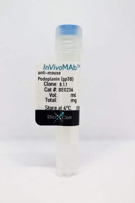
InVivoMAb anti-mouse Podoplanin (gp38)
Product Details
The 8.1.1 monoclonal antibody reacts with mouse podoplanin (PDPN) also known as glycoprotein 38 (gp38). Podoplanin is a 36 to 43 kDa mucin-type glycoprotein expressed by kidney glomerular epithelial cells (podocytes), lymphatic endothelial cells, and fibroblastic reticular cells. Podoplanin is the endogenous ligand for the C-type lectin receptor CLEC-2, which is expressed by platelets and DCs. CLEC-2 signaling is critical for platelet activation, the migration of activated DCs to draining lymph nodes, and maintenance of vascular integrity and lymph node structure. Podoplanin is critical for fibroblastic reticular cell contractility as well as during fetal development for blood-lymph separation and lung organogenesis. Podoplanin overexpression in cancer correlates with increased invasion and metastasis. The 8.1.1 antibody has been shown to block podoplanin in vivo and in vitro.Specifications
| Isotype | Syrian hamster IgG |
|---|---|
| Recommended Isotype Control(s) | InVivoMAb polyclonal Syrian hamster IgG |
| Recommended Dilution Buffer | InVivoPure pH 7.0 Dilution Buffer |
| Conjugation | This product is unconjugated. Conjugation is available via our Antibody Conjugation Services. |
| Immunogen | Mouse tymic epithelial cells |
| Reported Applications |
in vivo PDPN blockade in vitro PDPN blockade Immunofluorescence Western blot Flow cytometry |
| Formulation |
PBS, pH 7.0 Contains no stabilizers or preservatives |
| Endotoxin |
<2EU/mg (<0.002EU/μg) Determined by LAL gel clotting assay |
| Purity |
>95% Determined by SDS-PAGE |
| Sterility | 0.2 µm filtration |
| Production | Purified from cell culture supernatant in an animal-free facility |
| Purification | Protein A |
| RRID | AB_2687718 |
| Molecular Weight | 150 kDa |
| Storage | The antibody solution should be stored at the stock concentration at 4°C. Do not freeze. |
Recommended Products
-
Recommended Isotype Control(s)
InVivoMAb polyclonal Syrian hamster IgG
-

Recommended Dilution Buffer
InVivoPure pH 7.0 Dilution Buffer
Flow Cytometry
Rouhani, S. J., et al. (2015). "Roles of lymphatic endothelial cells expressing peripheral tissue antigens in CD4 T-cell tolerance induction" Nat Commun 6: 6771. PubMed
Lymphatic endothelial cells (LECs) directly express peripheral tissue antigens and induce CD8 T-cell deletional tolerance. LECs express MHC-II molecules, suggesting they might also tolerize CD4 T cells. We demonstrate that when beta-galactosidase (beta-gal) is expressed in LECs, beta-gal-specific CD8 T cells undergo deletion via the PD-1/PD-L1 and LAG-3/MHC-II pathways. In contrast, LECs do not present endogenous beta-gal in the context of MHC-II molecules to beta-gal-specific CD4 T cells. Lack of presentation is independent of antigen localization, as membrane-bound haemagglutinin and I-Ealpha are also not presented by MHC-II molecules. LECs express invariant chain and cathepsin L, but not H2-M, suggesting that they cannot load endogenous antigenic peptides onto MHC-II molecules. Importantly, LECs transfer beta-gal to dendritic cells, which subsequently present it to induce CD4 T-cell anergy. Therefore, LECs serve as an antigen reservoir for CD4 T-cell tolerance, and MHC-II molecules on LECs are used to induce CD8 T-cell tolerance via LAG-3.
in vivo PDPN blockade, in vitro PDPN blockade, Immunofluorescence, Flow Cytometry
Astarita, J. L., et al. (2015). "The CLEC-2-podoplanin axis controls the contractility of fibroblastic reticular cells and lymph node microarchitecture" Nat Immunol 16(1): 75-84. PubMed
In lymph nodes, fibroblastic reticular cells (FRCs) form a collagen-based reticular network that supports migratory dendritic cells (DCs) and T cells and transports lymph. A hallmark of FRCs is their propensity to contract collagen, yet this function is poorly understood. Here we demonstrate that podoplanin (PDPN) regulates actomyosin contractility in FRCs. Under resting conditions, when FRCs are unlikely to encounter mature DCs expressing the PDPN receptor CLEC-2, PDPN endowed FRCs with contractile function and exerted tension within the reticulum. Upon inflammation, CLEC-2 on mature DCs potently attenuated PDPN-mediated contractility, which resulted in FRC relaxation and reduced tissue stiffness. Disrupting PDPN function altered the homeostasis and spacing of FRCs and T cells, which resulted in an expanded reticular network and enhanced immunity.
Flow Cytometry
Fleige, H., et al. (2014). "IL-17-induced CXCL12 recruits B cells and induces follicle formation in BALT in the absence of differentiated FDCs" J Exp Med 211(4): 643-651. PubMed
Ectopic lymphoid tissue, such as bronchus-associated lymphoid tissue (BALT) in the lung, develops spontaneously at sites of chronic inflammation or during infection. The molecular mechanisms underlying the neogenesis of such tertiary lymphoid tissue are still poorly understood. We show that the type of inflammation-inducing pathogen determines which key factors are required for the formation and maturation of BALT. Thus, a single intranasal administration of the poxvirus modified vaccinia virus Ankara (MVA) is sufficient to induce highly organized BALT with densely packed B cell follicles containing a network of CXCL13-expressing follicular DCs (FDCs), as well as CXCL12-producing follicular stromal cells. In contrast, mice treated with P. aeruginosa (P.a.) develop BALT but B cell follicles lack FDCs while still harboring CXCL12-positive follicular stromal cells. Furthermore, in IL-17-deficient mice, P.a.-induced BALT largely lacks B cells as well as CXCL12-expressing stromal cells, and only loose infiltrates of T cells are present. We show that Toll-like receptor pathways are required for BALT induction by P.a., but not MVA, and provide evidence that IL-17 drives the differentiation of lung stroma toward podoplanin-positive CXCL12-expressing cells that allow follicle formation even in the absence of FDCs. Taken together, our results identify distinct pathogen-dependent induction and maturation pathways for BALT formation.
Flow Cytometry
Dubrot, J., et al. (2014). "Lymph node stromal cells acquire peptide-MHCII complexes from dendritic cells and induce antigen-specific CD4(+) T cell tolerance" J Exp Med 211(6): 1153-1166. PubMed
Dendritic cells (DCs), and more recently lymph node stromal cells (LNSCs), have been described to tolerize self-reactive CD8(+) T cells in LNs. Although LNSCs express MHCII, it is unknown whether they can also impact CD4(+) T cell functions. We show that the promoter IV (pIV) of class II transactivator (CIITA), the master regulator of MHCII expression, controls endogenous MHCII expression by LNSCs. Unexpectedly, LNSCs also acquire peptide-MHCII complexes from DCs and induce CD4(+) T cell dysfunction by presenting transferred complexes to naive CD4(+) T cells and preventing their proliferation and survival. Our data reveals a novel, alternative mechanism where LN-resident stromal cells tolerize CD4(+) T cells through the presentation of self-antigens via transferred peptide-MHCII complexes of DC origin.
Western Blot, Flow Cytometry
Pan, Y., et al. (2014). "Podoplanin requires sialylated O-glycans for stable expression on lymphatic endothelial cells and for interaction with platelets" Blood 124(24): 3656-3665. PubMed
O-glycosylation of podoplanin (PDPN) on lymphatic endothelial cells is critical for the separation of blood and lymphatic systems by interacting with platelet C-type lectin-like receptor 2 during development. However, how O-glycosylation controls endothelial PDPN function and expression remains unclear. In this study, we report that core 1 O-glycan-deficient or desialylated PDPN was highly susceptible to proteolytic degradation by various proteases, including metalloproteinases (MMP)-2/9. We found that the lymph contained activated MMP-2/9 and incubation of the lymph reduced surface levels of PDPN on core 1 O-glycan-deficient endothelial cells, but not on wild-type ECs. The lymph from mice with sepsis induced by cecal ligation and puncture, which contained bacteria-derived sialidase, reduced PDPN levels on wild-type ECs. The MMP inhibitor, GM6001, rescued these reductions. Additionally, GM6001 treatment rescued the reduction of PDPN level on lymphatic endothelial cells in mice lacking endothelial core 1 O-glycan or cecal ligation and puncture-treated mice. Furthermore, core 1 O-glycan-deficient or desialylated PDPN impaired platelet interaction under physiological flow. These data indicate that sialylated O-glycans of PDPN are essential for platelet adhesion and prevent PDPN from proteolytic degradation primarily mediated by MMPs in the lymph.
Immunofluorescence, Western Blot
Herzog, B. H., et al. (2013). "Podoplanin maintains high endothelial venule integrity by interacting with platelet CLEC-2" Nature 502(7469): 105-109. PubMed
Circulating lymphocytes continuously enter lymph nodes for immune surveillance through specialized blood vessels named high endothelial venules, a process that increases markedly during immune responses. How high endothelial venules (HEVs) permit lymphocyte transmigration while maintaining vascular integrity is unknown. Here we report a role for the transmembrane O-glycoprotein podoplanin (PDPN, also known as gp38 and T1alpha) in maintaining HEV barrier function. Mice with postnatal deletion of Pdpn lost HEV integrity and exhibited spontaneous bleeding in mucosal lymph nodes, and bleeding in the draining peripheral lymph nodes after immunization. Blocking lymphocyte homing rescued bleeding, indicating that PDPN is required to protect the barrier function of HEVs during lymphocyte trafficking. Further analyses demonstrated that PDPN expressed on fibroblastic reticular cells, which surround HEVs, functions as an activating ligand for platelet C-type lectin-like receptor 2 (CLEC-2, also known as CLEC1B). Mice lacking fibroblastic reticular cell PDPN or platelet CLEC-2 exhibited significantly reduced levels of VE-cadherin (also known as CDH5), which is essential for overall vascular integrity, on HEVs. Infusion of wild-type platelets restored HEV integrity in Clec-2-deficient mice. Activation of CLEC-2 induced release of sphingosine-1-phosphate from platelets, which promoted expression of VE-cadherin on HEVs ex vivo. Furthermore, draining peripheral lymph nodes of immunized mice lacking sphingosine-1-phosphate had impaired HEV integrity similar to Pdpn- and Clec-2-deficient mice. These data demonstrate that local sphingosine-1-phosphate release after PDPN-CLEC-2-mediated platelet activation is critical for HEV integrity during immune responses.
Chitinase 3-like-1 contributes to acetaminophen-induced liver injury by promoting hepatic platelet recruitment.
In eLife on 10 June 2021 by Shan, Z., Li, L., et al.
PubMed
Hepatic platelet accumulation contributes to acetaminophen (APAP)-induced liver injury (AILI). However, little is known about the molecular pathways involved in platelet recruitment to the liver and whether targeting such pathways could attenuate AILI. Mice were fasted overnight before intraperitoneally (i.p.) injected with APAP at a dose of 210 mg/kg for male mice and 325 mg/kg for female mice. Platelets adherent to Kupffer cells were determined in both mice and patients overdosed with APAP. The impact of α-chitinase 3-like-1 (α-Chi3l1) on alleviation of AILI was determined in a therapeutic setting, and liver injury was analyzed. The present study unveiled a critical role of Chi3l1 in hepatic platelet recruitment during AILI. Increased Chi3l1 and platelets in the liver were observed in patients and mice overdosed with APAP. Compared to wild-type (WT) mice, Chil1-/- mice developed attenuated AILI with markedly reduced hepatic platelet accumulation. Mechanistic studies revealed that Chi3l1 signaled through CD44 on macrophages to induce podoplanin expression, which mediated platelet recruitment through C-type lectin-like receptor 2. Moreover, APAP treatment of Cd44-/- mice resulted in much lower numbers of hepatic platelets and liver injury than WT mice, a phenotype similar to that in Chil1-/- mice. Recombinant Chi3l1 could restore hepatic platelet accumulation and AILI in Chil1-/- mice, but not in Cd44-/- mice. Importantly, we generated anti-Chi3l1 monoclonal antibodies and demonstrated that they could effectively inhibit hepatic platelet accumulation and AILI. We uncovered the Chi3l1/CD44 axis as a critical pathway mediating APAP-induced hepatic platelet recruitment and tissue injury. We demonstrated the feasibility and potential of targeting Chi3l1 to treat AILI. ZS received funding from NSFC (32071129). FWL received funding from NIH (GM123261). ALFSG received funding from NIDDK (DK 058369). ZA received funding from CPRIT (RP150551 and RP190561) and the Welch Foundation (AU-0042-20030616). CJ received funding from NIH (DK122708, DK109574, DK121330, and DK122796) and support from a University of Texas System Translational STARs award. Portions of this work were supported with resources and the use of facilities of the Michael E. DeBakey VA Medical Center and funding from Department of Veterans Affairs I01 BX002551 (Equipment, Personnel, Supplies). The contents do not represent the views of the US Department of Veterans Affairs or the US Government.
Chitinase 3-like-1 Contributes to Acetaminophen-induced Liver Injury by Promoting Hepatic Platelet Recruitment
Preprint on BioRxiv : the Preprint Server for Biology on 9 April 2021 by Shan, Z., Li, L., et al.
PubMed
Hepatic platelet accumulation contributes to acetaminophen (APAP)-induced liver injury (AILI). However, little is known about the molecular pathways involved in platelet recruitment to the liver and whether targeting such pathways could attenuate AILI. The present study unveiled a critical role of chitinase 3-like-1 (Chi3l1) in hepatic platelet recruitment during AILI. Increased Chi3l1 and platelets in the liver were observed in patients and mice overdosed with APAP. Compared to wild-type (WT) mice, Chi3l1 -/- mice developed attenuated AILI with markedly reduced hepatic platelet accumulation. Mechanistic studies revealed that Chi3l1 signaled through CD44 on macrophages to induce podoplanin expression, which mediated platelet recruitment through C-type lectin-like receptor 2. Moreover, APAP treatment of CD44 -/- mice resulted in much lower numbers of hepatic platelets and liver injury than WT mice, a phenotype similar to that in Chi3l1 -/- mice. Recombinant Chi3l1 could restore hepatic platelet accumulation and AILI in Chi3l1 -/- mice, but not in CD44 -/- mice. Importantly, we generated anti-Chi3l1 monoclonal antibodies and demonstrated that they could effectively inhibit hepatic platelet accumulation and AILI. Overall, we uncovered the Chi3l1/CD44 axis as a critical pathway mediating APAP-induced hepatic platelet recruitment and tissue injury. We demonstrated the feasibility and potential of targeting Chi3l1 to treat AILI.
- IHC-IF,
- FC/FACS,
- Homo sapiens (Human),
- Cancer Research,
- Pathology
Myeloid-Derived Lymphatic Endothelial Cell Progenitors Significantly Contribute to Lymphatic Metastasis in Clinical Breast Cancer.
In The American Journal of Pathology on 1 November 2019 by Volk-Draper, L., Patel, R., et al.
PubMed
Lymphatic metastasis is a high-impact prognostic factor for mortality of breast cancer (BC) patients, and it directly depends on tumor-associated lymphatic vessels. We previously reported that lipopolysaccharide-induced inflammatory lymphangiogenesis is strongly promoted by myeloid-derived lymphatic endothelial cell progenitors (M-LECPs) derived from the bone marrow (BM). As BC recruits massive numbers of provascular myeloid cells, we hypothesized that M-LECPs, within this recruited population, are specifically programmed to promote tumor lymphatics that increase lymph node metastasis. In support of this hypothesis, high levels of M-LECPs were found in peripheral blood and tumor tissues of BC patients. Moreover, the density of M-LECPs and lymphatic vessels positive for myeloid marker proteins strongly correlated with patient node status. It was also established that tumor M-LECPs coexpress lymphatic-specific, stem/progenitor and M2-type macrophage markers that indicate their BM hematopoietic-myeloid origin and distinguish them from mature lymphatic endothelial cells, tumor-infiltrating lymphoid cells, and tissue-resident macrophages. Using four orthotopic BC models, we show that mouse M-LECPs are similarly recruited to tumors and integrate into preexisting lymphatics. Finally, we demonstrate that adoptive transfer of in vitro differentiated M-LECPs, but not naïve or nondifferentiated BM cells, significantly increased metastatic burden in ipsilateral lymph nodes. These data support a causative role of BC-induced lymphatic progenitors in tumor lymphangiogenesis and suggest molecular targets for their inhibition. Copyright © 2019 American Society for Investigative Pathology. Published by Elsevier Inc. All rights reserved.