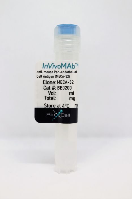
InVivoMAb anti-mouse Pan-endothelial Cell Antigen (MECA-32)
Product Details
The MECA-32 monoclonal antibody reacts with mouse pan-endothelial cell antigen also known as MECA-32 and PLVAP (plasmalemma vesicle-associated protein). MECA-32 is a 60 kDa transmembrane homodimer which is expressed on the surface of most endothelial cells. However, MECA-32 shows restricted distribution in the skeletal, cardiac and brain tissues of adult and embryonic mice. In embryonic mice, MECA-32 expression is developmentally regulated in the vessels associated with the blood brain barrier and persists only through day 16 of gestation.Specifications
| Isotype | Rat IgG2a, κ |
|---|---|
| Recommended Isotype Control(s) | InVivoMAb rat IgG2a isotype control, anti-trinitrophenol |
| Recommended Dilution Buffer | InVivoPure pH 7.0 Dilution Buffer |
| Conjugation | This product is unconjugated. Conjugation is available via our Antibody Conjugation Services. |
| Immunogen | Mouse lymph node stromal cells |
| Reported Applications |
Immunohistochemistry (frozen) Flow cytometry Western blot Immunofluorescence |
| Formulation |
PBS, pH 7.0 Contains no stabilizers or preservatives |
| Endotoxin |
<2EU/mg (<0.002EU/μg) Determined by LAL gel clotting assay |
| Purity |
>95% Determined by SDS-PAGE |
| Sterility | 0.2 µm filtration |
| Production | Purified from cell culture supernatant in an animal-free facility |
| Purification | Protein G |
| RRID | AB_10950167 |
| Molecular Weight | 150 kDa |
| Storage | The antibody solution should be stored at the stock concentration at 4°C. Do not freeze. |
Recommended Products
Immunofluorescence, Western Blot
Rantakari, P., et al. (2016). "Fetal liver endothelium regulates the seeding of tissue-resident macrophages" Nature 538(7625): 392-396. PubMed
Macrophages are required for normal embryogenesis, tissue homeostasis and immunity against microorganisms and tumours. Adult tissue-resident macrophages largely originate from long-lived, self-renewing embryonic precursors and not from haematopoietic stem-cell activity in the bone marrow. Although fate-mapping studies have uncovered a great amount of detail on the origin and kinetics of fetal macrophage development in the yolk sac and liver, the molecules that govern the tissue-specific migration of these cells remain completely unknown. Here we show that an endothelium-specific molecule, plasmalemma vesicle-associated protein (PLVAP), regulates the seeding of fetal monocyte-derived macrophages to tissues in mice. We found that PLVAP-deficient mice have completely normal levels of both yolk-sac- and bone-marrow-derived macrophages, but that fetal liver monocyte-derived macrophage populations were practically missing from tissues. Adult PLVAP-deficient mice show major alterations in macrophage-dependent iron recycling and mammary branching morphogenesis. PLVAP forms diaphragms in the fenestrae of liver sinusoidal endothelium during embryogenesis, interacts with chemoattractants and adhesion molecules and regulates the egress of fetal liver monocytes to the systemic vasculature. Thus, PLVAP selectively controls the exit of macrophage precursors from the fetal liver and, to our knowledge, is the first molecule identified in any organ as regulating the migratory events during embryonic macrophage ontogeny.
Immunohistochemistry (frozen)
Volk-Draper, L., et al. (2014). "Paclitaxel therapy promotes breast cancer metastasis in a TLR4-dependent manner" Cancer Res 74(19): 5421-5434. PubMed
Emerging evidence suggests that cytotoxic therapy may actually promote drug resistance and metastasis while inhibiting the growth of primary tumors. Work in preclinical models of breast cancer has shown that acquired chemoresistance to the widely used drug paclitaxel can be mediated by activation of the Toll-like receptor TLR4 in cancer cells. In this study, we determined the prometastatic effects of tumor-expressed TLR4 and paclitaxel therapy and investigated the mechanisms mediating these effects. While paclitaxel treatment was largely efficacious in inhibiting TLR4-negative tumors, it significantly increased the incidence and burden of pulmonary and lymphatic metastasis by TLR4-positive tumors. TLR4 activation by paclitaxel strongly increased the expression of inflammatory mediators, not only locally in the primary tumor microenvironment but also systemically in the blood, lymph nodes, spleen, bone marrow, and lungs. These proinflammatory changes promoted the outgrowth of Ly6C(+) and Ly6G(+) myeloid progenitor cells and their mobilization to tumors, where they increased blood vessel formation but not invasion of these vessels. In contrast, paclitaxel-mediated activation of TLR4-positive tumors induced de novo generation of deep intratumoral lymphatic vessels that were highly permissive to invasion by malignant cells. These results suggest that paclitaxel therapy of patients with TLR4-expressing tumors may activate systemic inflammatory circuits that promote angiogenesis, lymphangiogenesis, and metastasis, both at local sites and premetastatic niches where invasion occurs in distal organs. Taken together, our findings suggest that efforts to target TLR4 on tumor cells may simultaneously quell local and systemic inflammatory pathways that promote malignant progression, with implications for how to prevent tumor recurrence and the establishment of metastatic lesions, either during chemotherapy or after it is completed.
Flow Cytometry
Tkachenko, E., et al. (2012). "Caveolae, fenestrae and transendothelial channels retain PV1 on the surface of endothelial cells" PLoS One 7(3): e32655. PubMed
PV1 protein is an essential component of stomatal and fenestral diaphragms, which are formed at the plasma membrane of endothelial cells (ECs), on structures such as caveolae, fenestrae and transendothelial channels. Knockout of PV1 in mice results in in utero and perinatal mortality. To be able to interpret the complex PV1 knockout phenotype, it is critical to determine whether the formation of diaphragms is the only cellular role of PV1. We addressed this question by measuring the effect of complete and partial removal of structures capable of forming diaphragms on PV1 protein level. Removal of caveolae in mice by knocking out caveolin-1 or cavin-1 resulted in a dramatic reduction of PV1 protein level in lungs but not kidneys. The magnitude of PV1 reduction correlated with the abundance of structures capable of forming diaphragms in the microvasculature of these organs. The absence of caveolae in the lung ECs did not affect the transcription or translation of PV1, but it caused a sharp increase in PV1 protein internalization rate via a clathrin- and dynamin-independent pathway followed by degradation in lysosomes. Thus, PV1 is retained on the cell surface of ECs by structures capable of forming diaphragms, but undergoes rapid internalization and degradation in the absence of these structures, suggesting that formation of diaphragms is the only role of PV1.
- IHC-IF,
- Mus musculus (House mouse),
- Immunology and Microbiology
Blocking P2X7 receptor with AZ 10606120 exacerbates vascular hyperpermeability and inflammation in murine polymicrobial sepsis.
In Physiological Reports on 1 June 2022 by Meegan, J. E., Komalavilas, P., et al.
PubMed
Sepsis is a devastating disease with high morbidity and mortality and no specific treatments. The pathophysiology of sepsis involves a hyperinflammatory response and release of damage-associated molecular patterns (DAMPs), including adenosine triphosphate (ATP), from activated and dying cells. Purinergic receptors activated by ATP have gained attention for their roles in sepsis, which can be pro- or anti-inflammatory depending on the context. Current data regarding the role of ATP-specific purinergic receptor P2X7 (P2X7R) in vascular function and inflammation during sepsis are conflicting, and its role on the endothelium has not been well characterized. In this study, we hypothesized that the P2X7R antagonist AZ 10606120 (AZ106) would prevent endothelial dysfunction during sepsis. As proof of concept, we first demonstrated the ability of AZ106 (10 µM) to prevent endothelial dysfunction in intact rat aorta in response to IL-1β, an inflammatory mediator upregulated during sepsis. Likewise, blocking P2X7R with AZ106 (10 µg/g) reduced the impairment of endothelial-dependent relaxation in mice subjected to intraperitoneal injection of cecal slurry (CS), a model of polymicrobial sepsis. However, contrary to our hypothesis, AZ106 did not improve microvascular permeability or injury, lung apoptosis, or illness severity in mice subjected to CS. Instead, AZ106 elevated spleen bacterial burden and circulating inflammatory markers. In conclusion, antagonism of P2X7R signaling during sepsis appears to disrupt the balance between its roles in inflammatory, antimicrobial, and vascular function. © 2022 The Authors. Physiological Reports published by Wiley Periodicals LLC on behalf of The Physiological Society and the American Physiological Society.
- IF,
- IHC-IF,
- Mus musculus (House mouse)
Fenestral diaphragms and PLVAP associations in liver sinusoidal endothelial cells are developmentally regulated.
In Scientific Reports on 30 October 2019 by Auvinen, K., Lokka, E., et al.
PubMed
Endothelial cells contain several nanoscale domains such as caveolae, fenestrations and transendothelial channels, which regulate signaling and transendothelial permeability. These structures can be covered by filter-like diaphragms. A transmembrane PLVAP (plasmalemma vesicle associated protein) protein has been shown to be necessary for the formation of diaphragms. The expression, subcellular localization and fenestra-forming role of PLVAP in liver sinusoidal endothelial cells (LSEC) have remained controversial. Here we show that fenestrations in LSEC contain PLVAP-diaphragms during the fetal angiogenesis, but they lose the diaphragms at birth. Although it is thought that PLVAP only localizes to diaphragms, we found luminal localization of PLVAP in adult LSEC using several imaging techniques. Plvap-deficient mice revealed that the absence of PLVAP and diaphragms did not affect the morphology, the number of fenestrations or the overall vascular architecture in the liver sinusoids. Nevertheless, PLVAP in fetal LSEC (fenestrations with diaphragms) associated with LYVE-1 (lymphatic vessel endothelial hyaluronan receptor 1), neuropilin-1 and VEGFR2 (vascular endothelial growth factor receptor 2), whereas in the adult LSEC (fenestrations without diaphragms) these complexes disappeared. Collectively, our data show that PLVAP can be expressed on endothelial cells without diaphragms, contradict the prevailing concept that biogenesis of fenestrae would be PLVAP-dependent, and reveal previously unknown PLVAP-dependent molecular complexes in LSEC during angiogenesis.
- Mus musculus (House mouse)
Integrated Brain Atlas for Unbiased Mapping of Nervous System Effects Following Liraglutide Treatment.
In Scientific Reports on 9 July 2018 by Salinas, C. B. G., Lu, T. T., et al.
PubMed
Light Sheet Fluorescence Microscopy (LSFM) of whole organs, in particular the brain, offers a plethora of biological data imaged in 3D. This technique is however often hindered by cumbersome non-automated analysis methods. Here we describe an approach to fully automate the analysis by integrating with data from the Allen Institute of Brain Science (AIBS), to provide precise assessment of the distribution and action of peptide-based pharmaceuticals in the brain. To illustrate this approach, we examined the acute central nervous system effects of the glucagon-like peptide-1 (GLP-1) receptor agonist liraglutide. Peripherally administered liraglutide accessed the hypothalamus and brainstem, and led to activation in several brain regions of which most were intersected by projections from neurons in the lateral parabrachial nucleus. Collectively, we provide a rapid and unbiased analytical framework for LSFM data which enables quantification and exploration based on data from AIBS to support basic and translational discovery.
Disrupting the Btk Pathway Suppresses COPD-Like Lung Alterations in Atherosclerosis Prone ApoE-/- Mice Following Regular Exposure to Cigarette Smoke.
In International Journal of Molecular Sciences on 24 January 2018 by Florence, J. M., Krupa, A., et al.
PubMed
Chronic obstructive pulmonary disease (COPD) is associated with severe chronic inflammation that promotes irreversible tissue destruction. Moreover, the most broadly accepted cause of COPD is exposure to cigarette smoke. There is no effective cure and significantly, the mechanism behind the development and progression of this disease remains unknown. Our laboratory has demonstrated that Bruton's tyrosine kinase (Btk) is a critical regulator of pro-inflammatory processes in the lungs and that Btk controls expression of matrix metalloproteinase-9 (MMP-9) in the alveolar compartment. For this study apolipoprotein E null (ApoE-/-) mice were exposed to SHS to facilitate study in a COPD/atherosclerosis comorbidity model. We applied two types of treatments, animals received either a pharmacological inhibitor of Btk or MMP-9 specific siRNA to minimize MMP-9 expression in endothelial cells or neutrophils. We have shown that these treatments had a protective effect in the lung. We have noted a decrease in alveolar changes related to SHS induced inflammation in treated animals. In summary, we are presenting a novel concept in the field of COPD, i.e., that Btk may be a new drug target for this disease. Moreover, cell specific targeting of MMP-9 may also benefit patients affected by this disease.
- Cancer Research
Paclitaxel therapy promotes breast cancer metastasis in a TLR4-dependent manner.
In Cancer Research on 1 October 2014 by Volk-Draper, L., Hall, K., et al.
PubMed
Emerging evidence suggests that cytotoxic therapy may actually promote drug resistance and metastasis while inhibiting the growth of primary tumors. Work in preclinical models of breast cancer has shown that acquired chemoresistance to the widely used drug paclitaxel can be mediated by activation of the Toll-like receptor TLR4 in cancer cells. In this study, we determined the prometastatic effects of tumor-expressed TLR4 and paclitaxel therapy and investigated the mechanisms mediating these effects. While paclitaxel treatment was largely efficacious in inhibiting TLR4-negative tumors, it significantly increased the incidence and burden of pulmonary and lymphatic metastasis by TLR4-positive tumors. TLR4 activation by paclitaxel strongly increased the expression of inflammatory mediators, not only locally in the primary tumor microenvironment but also systemically in the blood, lymph nodes, spleen, bone marrow, and lungs. These proinflammatory changes promoted the outgrowth of Ly6C(+) and Ly6G(+) myeloid progenitor cells and their mobilization to tumors, where they increased blood vessel formation but not invasion of these vessels. In contrast, paclitaxel-mediated activation of TLR4-positive tumors induced de novo generation of deep intratumoral lymphatic vessels that were highly permissive to invasion by malignant cells. These results suggest that paclitaxel therapy of patients with TLR4-expressing tumors may activate systemic inflammatory circuits that promote angiogenesis, lymphangiogenesis, and metastasis, both at local sites and premetastatic niches where invasion occurs in distal organs. Taken together, our findings suggest that efforts to target TLR4 on tumor cells may simultaneously quell local and systemic inflammatory pathways that promote malignant progression, with implications for how to prevent tumor recurrence and the establishment of metastatic lesions, either during chemotherapy or after it is completed. ©2014 American Association for Cancer Research.


