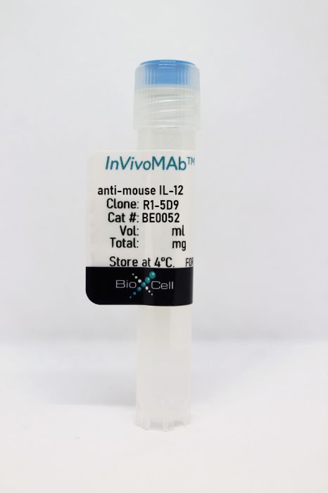
InVivoMAb anti-mouse IL-12
Product Details
The R1-5D9 antibody reacts with mouse IL-12. IL-12 is a heterodimeric cytokine composed of subunits IL-12α p35 and IL-12β p40. IL-12 is secreted by activated monocytes, macrophages, and dendritic cells. IL-12 plays roles in T lymphocyte differentiation, IFNγ production, and NK cell cytotoxicity. Overexpression of IL-12 p40 was observed in the central nervous system of patients with multiple sclerosis, suggesting a role of this cytokine in the pathogenesis of the disease.Specifications
| Isotype | Rat IgG2a |
|---|---|
| Recommended Isotype Control(s) | InVivoMAb rat IgG2a isotype control, anti-trinitrophenol |
| Recommended Dilution Buffer | InVivoPure pH 7.0 Dilution Buffer |
| Conjugation | This product is unconjugated. Conjugation is available via our Antibody Conjugation Services. |
| Immunogen | Recombinant mouse IL-12 p75 |
| Reported Applications |
in vivo IL-12 neutralization in vitro IL-12 neutralization |
| Formulation |
PBS, pH 7.0 Contains no stabilizers or preservatives |
| Endotoxin |
<2EU/mg (<0.002EU/μg) Determined by LAL gel clotting assay |
| Purity |
>95% Determined by SDS-PAGE |
| Sterility | 0.2 µm filtration |
| Production | Purified from cell culture supernatant in an animal-free facility |
| Purification | Protein G |
| RRID | AB_1107700 |
| Molecular Weight | 150 kDa |
| Storage | The antibody solution should be stored at the stock concentration at 4°C. Do not freeze. |
Recommended Products
in vivo IL-12 neutralization
Gimblet, C., et al. (2017). "Cutaneous Leishmaniasis Induces a Transmissible Dysbiotic Skin Microbiota that Promotes Skin Inflammation" Cell Host Microbe 22(1): 13-24.e14. PubMed
Skin microbiota can impact allergic and autoimmune responses, wound healing, and anti-microbial defense. We investigated the role of skin microbiota in cutaneous leishmaniasis and found that human patients infected with Leishmania braziliensis develop dysbiotic skin microbiota, characterized by increases in the abundance of Staphylococcus and/or Streptococcus. Mice infected with L. major exhibit similar changes depending upon disease severity. Importantly, this dysbiosis is not limited to the lesion site, but is transmissible to normal skin distant from the infection site and to skin from co-housed naive mice. This observation allowed us to test whether a pre-existing dysbiotic skin microbiota influences disease, and we found that challenging dysbiotic naive mice with L. major or testing for contact hypersensitivity results in exacerbated skin inflammatory responses. These findings demonstrate that a dysbiotic skin microbiota is not only a consequence of tissue stress, but also enhances inflammation, which has implications for many inflammatory cutaneous diseases.
in vitro IL-12 neutralization
Choi, Y. S., et al. (2015). "LEF-1 and TCF-1 orchestrate TFH differentiation by regulating differentiation circuits upstream of the transcriptional repressor Bcl6" Nat Immunol 16(9): 980-990. PubMed
Follicular helper T cells (TFH cells) are specialized effector CD4(+) T cells that help B cells develop germinal centers (GCs) and memory. However, the transcription factors that regulate the differentiation of TFH cells remain incompletely understood. Here we report that selective loss of Lef1 or Tcf7 (which encode the transcription factor LEF-1 or TCF-1, respectively) resulted in TFH cell defects, while deletion of both Lef1 and Tcf7 severely impaired the differentiation of TFH cells and the formation of GCs. Forced expression of LEF-1 enhanced TFH differentiation. LEF-1 and TCF-1 coordinated such differentiation by two general mechanisms. First, they established the responsiveness of naive CD4(+) T cells to TFH cell signals. Second, they promoted early TFH differentiation via the multipronged approach of sustaining expression of the cytokine receptors IL-6Ralpha and gp130, enhancing expression of the costimulatory receptor ICOS and promoting expression of the transcriptional repressor Bcl6.
in vitro IL-12 neutralization
Bertin, S., et al. (2014). "The ion channel TRPV1 regulates the activation and proinflammatory properties of CD4(+) T cells" Nat Immunol 15(11): 1055-1063. PubMed
TRPV1 is a Ca(2+)-permeable channel studied mostly as a pain receptor in sensory neurons. However, its role in other cell types is poorly understood. Here we found that TRPV1 was functionally expressed in CD4(+) T cells, where it acted as a non-store-operated Ca(2+) channel and contributed to T cell antigen receptor (TCR)-induced Ca(2+) influx, TCR signaling and T cell activation. In models of T cell-mediated colitis, TRPV1 promoted colitogenic T cell responses and intestinal inflammation. Furthermore, genetic and pharmacological inhibition of TRPV1 in human CD4(+) T cells recapitulated the phenotype of mouse Trpv1(-/-) CD4(+) T cells. Our findings suggest that inhibition of TRPV1 could represent a new therapeutic strategy for restraining proinflammatory T cell responses.
- In Vivo,
- Mus musculus (House mouse),
- Immunology and Microbiology
Commensal Cryptosporidium colonization elicits a cDC1-dependent Th1 response that promotes intestinal homeostasis and limits other infections.
In Immunity on 9 November 2021 by Russler-Germain, E. V., Jung, J., et al.
PubMed
Cryptosporidium can cause severe diarrhea and morbidity, but many infections are asymptomatic. Here, we studied the immune response to a commensal strain of Cryptosporidium tyzzeri (Ct-STL) serendipitously discovered when conventional type 1 dendritic cell (cDC1)-deficient mice developed cryptosporidiosis. Ct-STL was vertically transmitted without negative health effects in wild-type mice. Yet, Ct-STL provoked profound changes in the intestinal immune system, including induction of an IFN-γ-producing Th1 response. TCR sequencing coupled with in vitro and in vivo analysis of common Th1 TCRs revealed that Ct-STL elicited a dominant antigen-specific Th1 response. In contrast, deficiency in cDC1s skewed the Ct-STL CD4 T cell response toward Th17 and regulatory T cells. Although Ct-STL predominantly colonized the small intestine, colon Th1 responses were enhanced and associated with protection against Citrobacter rodentium infection and exacerbation of dextran sodium sulfate and anti-IL10R-triggered colitis. Thus, Ct-STL represents a commensal pathobiont that elicits Th1-mediated intestinal homeostasis that may reflect asymptomatic human Cryptosporidium infection. Copyright © 2021 Elsevier Inc. All rights reserved.
- In Vitro,
- Mus musculus (House mouse),
- Immunology and Microbiology
Gut Helicobacter presentation by multiple dendritic cell subsets enables context-specific regulatory T cell generation.
In eLife on 3 February 2021 by Russler-Germain, E. V., Yi, J., et al.
PubMed
Generation of tolerogenic peripheral regulatory T (pTreg) cells is commonly thought to involve CD103+ gut dendritic cells (DCs), yet their role in commensal-reactive pTreg development is unclear. Using two Helicobacter-specific T cell receptor (TCR) transgenic mouse lines, we found that both CD103+ and CD103- migratory, but not resident, DCs from the colon-draining mesenteric lymph node presented Helicobacter antigens to T cells ex vivo. Loss of most CD103+ migratory DCs in vivo using murine genetic models did not affect the frequency of Helicobacter-specific pTreg cell generation or induce compensatory tolerogenic changes in the remaining CD103- DCs. By contrast, activation in a Th1-promoting niche in vivo blocked Helicobacter-specific pTreg generation. Thus, these data suggest a model where DC-mediated effector T cell differentiation is 'dominant', necessitating that all DC subsets presenting antigen are permissive for pTreg cell induction to maintain gut tolerance.
- Cell Culture,
- Mus musculus (House mouse)
Hectd3 promotes pathogenic Th17 lineage through Stat3 activation and Malt1 signaling in neuroinflammation.
In Nature Communications on 11 February 2019 by Cho, J. J., Xu, Z., et al.
PubMed
Polyubiquitination promotes proteasomal degradation, or signaling and localization, of targeted proteins. Here we show that the E3 ubiquitin ligase Hectd3 is necessary for pathogenic Th17 cell generation in experimental autoimmune encephalomyelitis (EAE), a mouse model for human multiple sclerosis. Hectd3-deficient mice have lower EAE severity, reduced Th17 program and inefficient Th17 cell differentiation. However, Stat3, but not RORγt, has decreased polyubiquitination, as well as diminished tyrosine-705 activating phosphorylation. Additionally, non-degradative polyubiquitination of Malt1, critical for NF-κB activation and Th17 cell function, is reduced. Mechanistically, Hectd3 promotes K27-linked and K29-linked polyubiquitin chains on Malt1, and K27-linked polyubiquitin chains on Stat3. Moreover, Stat3 K180 and Malt1 K648 are targeted by Hectd3 for non-degradative polyubiquitination to mediate robust generation of RORγt+IL-17Ahi effector CD4+ T cells. Thus, our studies delineate a mechanism connecting signaling related polyubiquitination of Malt1 and Stat3, leading to NF-kB activation and RORγt expression, to pathogenic Th17 cell function in EAE.
- Cell Culture,
- Mus musculus (House mouse),
- Immunology and Microbiology
Helicobacter species are potent drivers of colonic T cell responses in homeostasis and inflammation.
In Science Immunology on 21 July 2017 by Chai, J. N., Peng, Y., et al.
PubMed
Specific gut commensal bacteria improve host health by eliciting mutualistic regulatory T (Treg) cell responses. However, the bacteria that induce effector T (Teff) cells during inflammation are unclear. We addressed this by analyzing bacterial-reactive T cell receptor (TCR) transgenic cells and TCR repertoires in a murine colitis model. Unexpectedly, we found that mucosal-associated Helicobacter species triggered both Treg cell responses during homeostasis and Teff cell responses during colitis, as suggested by an increased overlap between the Teff/Treg TCR repertoires with colitis. Four of six Treg TCRs tested recognized mucosal-associated Helicobacter species in vitro and in vivo. By contrast, the marked expansion of luminal Bacteroides species seen during colitis did not trigger a commensurate Teff cell response. Unlike other Treg cell-inducing bacteria, Helicobacter species are known pathobionts and cause disease in immunodeficient mice. Thus, our study suggests a model in which mucosal bacteria elicit context-dependent Treg or Teff cell responses to facilitate intestinal tolerance or inflammation. Copyright © 2017 The Authors, some rights reserved; exclusive licensee American Association for the Advancement of Science. No claim to original U.S. Government Works.
- In Vivo,
- Mus musculus (House mouse),
- Immunology and Microbiology
Cutaneous Leishmaniasis Induces a Transmissible Dysbiotic Skin Microbiota that Promotes Skin Inflammation.
In Cell Host & Microbe on 12 July 2017 by Gimblet, C., Meisel, J. S., et al.
PubMed
Skin microbiota can impact allergic and autoimmune responses, wound healing, and anti-microbial defense. We investigated the role of skin microbiota in cutaneous leishmaniasis and found that human patients infected with Leishmania braziliensis develop dysbiotic skin microbiota, characterized by increases in the abundance of Staphylococcus and/or Streptococcus. Mice infected with L. major exhibit similar changes depending upon disease severity. Importantly, this dysbiosis is not limited to the lesion site, but is transmissible to normal skin distant from the infection site and to skin from co-housed naive mice. This observation allowed us to test whether a pre-existing dysbiotic skin microbiota influences disease, and we found that challenging dysbiotic naive mice with L. major or testing for contact hypersensitivity results in exacerbated skin inflammatory responses. These findings demonstrate that a dysbiotic skin microbiota is not only a consequence of tissue stress, but also enhances inflammation, which has implications for many inflammatory cutaneous diseases.Copyright © 2017 Elsevier Inc. All rights reserved.
- In Vivo,
- Mus musculus (House mouse),
- Immunology and Microbiology
The Costimulatory Molecule ICOS Regulates Host Th1 and Follicular Th Cell Differentiation in Response to Plasmodium chabaudi chabaudi AS Infection.
In The Journal of Immunology on 15 January 2016 by Wikenheiser, D. J., Ghosh, D., et al.
PubMed
Blood-stage Plasmodium chabaudi chabaudi AS infection requires cell- and Ab-mediated immunity to control acute and persistent infection, respectively. ICOS regulates CD4(+) T cell activation and promotes the induction of follicular Th (TFH) cells, CD4(+) T cells that support B cell affinity maturation within germinal centers (GCs), resulting in the production of high-affinity Abs. In this article, we demonstrate that, in response to P. c. chabaudi AS infection, the absence of ICOS resulted in an enhanced Th1 immune response that reduced peak parasitemia. Despite the absence of ICOS, CD4(+) T cells were capable of expressing PD-1, B cell lymphoma 6, and CXCR5 during early infection, indicating TFH development was not impaired. However, by day 21 postinfection, Icos(-/-) mice accumulated fewer splenic TFHs compared with Icos(+/+) mice, leading to substantially fewer GC B cells and a decrease in affinity, but not production, of parasite-specific isotype-switched Abs. Moreover, treatment of mice with anti-ICOS ligand Abs to modulate ICOS-ICOS ligand signaling revealed a requirement for ICOS in TFH differentiation only after day 6 postinfection. Ultimately, the quality and quantity of isotype-switched Abs produced in Icos(-/-) mice declined over time, resulting in impaired control of persistent parasitemia. Collectively, these data suggest ICOS is not required for TFH induction during P. c. chabaudi AS infection or production of isotype-switched Abs, but it is necessary for maintenance of a sustained high-affinity, protective Ab response. Copyright © 2016 by The American Association of Immunologists, Inc.


