InVivoMAb anti-mouse CD28
Product Details
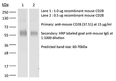
The 37.51 monoclonal antibody reacts with mouse CD28, a 45 kDa costimulatory receptor and a member of the Ig superfamily. CD28 is expressed by thymocytes, most peripheral T cells, and NK cells. CD28 is a receptor for CD80 (B7-1) and CD86 (B7-2). Signaling through CD28 augments IL-2 and IL-2 receptor expression as well as cytotoxicity of CD3-activated T cells. The 37.51 antibody has been shown to stimulate the proliferation and cytokine production by activated T and NK cells and provide a costimulatory signal for CTL induction.Specifications
| Isotype | Syrian Hamster IgG2 |
|---|---|
| Recommended Isotype Control(s) | InVivoMAb polyclonal Syrian hamster IgG |
| Recommended Dilution Buffer | InVivoPure pH 6.0T Dilution Buffer |
| Conjugation | This product is unconjugated. Conjugation is available via our Antibody Conjugation Services. |
| Immunogen | C57BL/6 mouse T cell lymphoma EL-4 cells |
| Reported Applications |
in vitro T cell stimulation/activation in vivo CD28 blockade |
| Formulation |
PBS, pH 6.0 0.01% Tween Contains no stabilizers or preservatives |
| Endotoxin |
<2EU/mg (<0.002EU/μg) Determined by LAL gel clotting assay |
| Purity |
>95% Determined by SDS-PAGE |
| Sterility | 0.2 µm filtration |
| Production | Purified from cell culture supernatant in an animal-free facility |
| Purification | Protein G |
| RRID | AB_1107624 |
| Molecular Weight | 150 kDa |
| Storage | The antibody solution should be stored at the stock concentration at 4°C. Do not freeze. |
Recommended Products
in vitro T cell stimulation/activation
Lacher, S. M., et al. (2018). "NF-kappaB inducing kinase (NIK) is an essential post-transcriptional regulator of T-cell activation affecting F-actin dynamics and TCR signaling" J Autoimmun 94: 110-121. PubMed
NF-kappaB inducing kinase (NIK) is the key protein of the non-canonical NF-kappaB pathway and is important for the development of lymph nodes and other secondary immune organs. We elucidated the specific role of NIK in T cells using T-cell specific NIK-deficient (NIK(DeltaT)) mice. Despite showing normal development of lymphoid organs, NIK(DeltaT) mice were resistant to induction of CNS autoimmunity. T cells from NIK(DeltaT) mice were deficient in late priming, failed to up-regulate T-bet and to transmigrate into the CNS. Proteomic analysis of activated NIK(-/-) T cells showed de-regulated expression of proteins involved in the formation of the immunological synapse: in particular, proteins involved in cytoskeleton dynamics. In line with this we found that NIK-deficient T cells were hampered in phosphorylation of Zap70, LAT, AKT, ERK1/2 and PLCgamma upon TCR engagement. Hence, our data disclose a hitherto unknown function of NIK in T-cell priming and differentiation.
in vitro T cell stimulation/activation
Wendland, K., et al. (2018). "Retinoic Acid Signaling in Thymic Epithelial Cells Regulates Thymopoiesis" J Immunol 201(2): 524-532. PubMed
Despite the essential role of thymic epithelial cells (TEC) in T cell development, the signals regulating TEC differentiation and homeostasis remain incompletely understood. In this study, we show a key in vivo role for the vitamin A metabolite, retinoic acid (RA), in TEC homeostasis. In the absence of RA signaling in TEC, cortical TEC (cTEC) and CD80(lo)MHC class II(lo) medullary TEC displayed subset-specific alterations in gene expression, which in cTEC included genes involved in epithelial proliferation, development, and differentiation. Mice whose TEC were unable to respond to RA showed increased cTEC proliferation, an accumulation of stem cell Ag-1(hi) cTEC, and, in early life, a decrease in medullary TEC numbers. These alterations resulted in reduced thymic cellularity in early life, a reduction in CD4 single-positive and CD8 single-positive numbers in both young and adult mice, and enhanced peripheral CD8(+) T cell survival upon TCR stimulation. Collectively, our results identify RA as a regulator of TEC homeostasis that is essential for TEC function and normal thymopoiesis.
in vitro T cell stimulation/activation
Ron-Harel, N., et al. (2016). "Mitochondrial Biogenesis and Proteome Remodeling Promote One-Carbon Metabolism for T Cell Activation" Cell Metab 24(1): 104-117. PubMed
Naive T cell stimulation activates anabolic metabolism to fuel the transition from quiescence to growth and proliferation. Here we show that naive CD4(+) T cell activation induces a unique program of mitochondrial biogenesis and remodeling. Using mass spectrometry, we quantified protein dynamics during T cell activation. We identified substantial remodeling of the mitochondrial proteome over the first 24 hr of T cell activation to generate mitochondria with a distinct metabolic signature, with one-carbon metabolism as the most induced pathway. Salvage pathways and mitochondrial one-carbon metabolism, fed by serine, contribute to purine and thymidine synthesis to enable T cell proliferation and survival. Genetic inhibition of the mitochondrial serine catabolic enzyme SHMT2 impaired T cell survival in culture and antigen-specific T cell abundance in vivo. Thus, during T cell activation, mitochondrial proteome remodeling generates specialized mitochondria with enhanced one-carbon metabolism that is critical for T cell activation and survival.
in vitro T cell stimulation/activation
Gu, A. D., et al. (2015). "A critical role for transcription factor Smad4 in T cell function that is independent of transforming growth factor beta receptor signaling" Immunity 42(1): 68-79. PubMed
Transforming growth factor-beta (TGF-beta) suppresses T cell function to maintain self-tolerance and to promote tumor immune evasion. Yet how Smad4, a transcription factor component of TGF-beta signaling, regulates T cell function remains unclear. Here we have demonstrated an essential role for Smad4 in promoting T cell function during autoimmunity and anti-tumor immunity. Smad4 deletion rescued the lethal autoimmunity resulting from transforming growth factor-beta receptor (TGF-betaR) deletion and compromised T-cell-mediated tumor rejection. Although Smad4 was dispensable for T cell generation, homeostasis, and effector function, it was essential for T cell proliferation after activation in vitro and in vivo. The transcription factor Myc was identified to mediate Smad4-controlled T cell proliferation. This study thus reveals a requirement of Smad4 for T-cell-mediated autoimmunity and tumor rejection, which is beyond the current paradigm. It highlights a TGF-betaR-independent role for Smad4 in promoting T cell function, autoimmunity, and anti-tumor immunity.
in vitro T cell stimulation/activation
Choi, Y. S., et al. (2015). "LEF-1 and TCF-1 orchestrate TFH differentiation by regulating differentiation circuits upstream of the transcriptional repressor Bcl6" Nat Immunol 16(9): 980-990. PubMed
Follicular helper T cells (TFH cells) are specialized effector CD4(+) T cells that help B cells develop germinal centers (GCs) and memory. However, the transcription factors that regulate the differentiation of TFH cells remain incompletely understood. Here we report that selective loss of Lef1 or Tcf7 (which encode the transcription factor LEF-1 or TCF-1, respectively) resulted in TFH cell defects, while deletion of both Lef1 and Tcf7 severely impaired the differentiation of TFH cells and the formation of GCs. Forced expression of LEF-1 enhanced TFH differentiation. LEF-1 and TCF-1 coordinated such differentiation by two general mechanisms. First, they established the responsiveness of naive CD4(+) T cells to TFH cell signals. Second, they promoted early TFH differentiation via the multipronged approach of sustaining expression of the cytokine receptors IL-6Ralpha and gp130, enhancing expression of the costimulatory receptor ICOS and promoting expression of the transcriptional repressor Bcl6.
in vitro T cell stimulation/activation
Xu, H., et al. (2015). "Regulation of bifurcating B cell trajectories by mutual antagonism between transcription factors IRF4 and IRF8" Nat Immunol . PubMed
Upon recognition of antigen, B cells undertake a bifurcated response in which some cells rapidly differentiate into plasmablasts while others undergo affinity maturation in germinal centers (GCs). Here we identified a double-negative feedback loop between the transcription factors IRF4 and IRF8 that regulated the initial developmental bifurcation of activated B cells as well as the GC response. IRF8 dampened signaling via the B cell antigen receptor (BCR), facilitated antigen-specific interaction with helper T cells, and promoted antibody affinity maturation while antagonizing IRF4-driven differentiation of plasmablasts. Genomic analysis revealed concentration-dependent actions of IRF4 and IRF8 in regulating distinct gene-expression programs. Stochastic modeling suggested that the double-negative feedback was sufficient to initiate bifurcation of the B cell developmental trajectories.
in vivo CD28 blockade
Rouhani, S. J., et al. (2015). "Roles of lymphatic endothelial cells expressing peripheral tissue antigens in CD4 T-cell tolerance induction" Nat Commun 6: 6771. PubMed
Lymphatic endothelial cells (LECs) directly express peripheral tissue antigens and induce CD8 T-cell deletional tolerance. LECs express MHC-II molecules, suggesting they might also tolerize CD4 T cells. We demonstrate that when beta-galactosidase (beta-gal) is expressed in LECs, beta-gal-specific CD8 T cells undergo deletion via the PD-1/PD-L1 and LAG-3/MHC-II pathways. In contrast, LECs do not present endogenous beta-gal in the context of MHC-II molecules to beta-gal-specific CD4 T cells. Lack of presentation is independent of antigen localization, as membrane-bound haemagglutinin and I-Ealpha are also not presented by MHC-II molecules. LECs express invariant chain and cathepsin L, but not H2-M, suggesting that they cannot load endogenous antigenic peptides onto MHC-II molecules. Importantly, LECs transfer beta-gal to dendritic cells, which subsequently present it to induce CD4 T-cell anergy. Therefore, LECs serve as an antigen reservoir for CD4 T-cell tolerance, and MHC-II molecules on LECs are used to induce CD8 T-cell tolerance via LAG-3.
in vitro T cell stimulation/activation
Rabenstein, H., et al. (2014). "Differential kinetics of antigen dependency of CD4+ and CD8+ T cells" J Immunol 192(8): 3507-3517. PubMed
Ag recognition via the TCR is necessary for the expansion of specific T cells that then contribute to adaptive immunity as effector and memory cells. Because CD4+ and CD8+ T cells differ in terms of their priming APCs and MHC ligands we compared their requirements of Ag persistence during their expansion phase side by side. Proliferation and effector differentiation of TCR transgenic and polyclonal mouse T cells were thus analyzed after transient and continuous TCR signals. Following equally strong stimulation, CD4+ T cell proliferation depended on prolonged Ag presence, whereas CD8+ T cells were able to divide and differentiate into effector cells despite discontinued Ag presentation. CD4+ T cell proliferation was neither affected by Th lineage or memory differentiation nor blocked by coinhibitory signals or missing inflammatory stimuli. Continued CD8+ T cell proliferation was truly independent of self-peptide/MHC-derived signals. The subset divergence was also illustrated by surprisingly broad transcriptional differences supporting a stronger propensity of CD8+ T cells to programmed expansion. These T cell data indicate an intrinsic difference between CD4+ and CD8+ T cells regarding the processing of TCR signals for proliferation. We also found that the presentation of a MHC class II-restricted peptide is more efficiently prolonged by dendritic cell activation in vivo than a class I bound one. In summary, our data demonstrate that CD4+ T cells require continuous stimulation for clonal expansion, whereas CD8+ T cells can divide following a much shorter TCR signal.
in vitro T cell stimulation/activation
Xiao, N., et al. (2014). "The E3 ubiquitin ligase Itch is required for the differentiation of follicular helper T cells" Nat Immunol 15(7): 657-666. PubMed
Follicular helper T cells (T(FH) cells) are responsible for effective B cell-mediated immunity, and Bcl-6 is a central factor for the differentiation of T(FH) cells. However, the molecular mechanisms that regulate the induction of T(FH) cells remain unclear. Here we found that the E3 ubiquitin ligase Itch was essential for the differentiation of T(FH) cells, germinal center responses and immunoglobulin G (IgG) responses to acute viral infection. Itch acted intrinsically in CD4(+) T cells at early stages of T(FH) cell development. Itch seemed to act upstream of Bcl-6 expression, as Bcl-6 expression was substantially impaired in Itch(-/-) cells, and the differentiation of Itch(-/-) T cells into T(FH) cells was restored by enforced expression of Bcl-6. Itch associated with the transcription factor Foxo1 and promoted its ubiquitination and degradation. The defective T(FH) differentiation of Itch(-/-) T cells was rectified by deletion of Foxo1. Thus, our results indicate that Itch acts as an essential positive regulator in the differentiation of T(FH) cells.
in vitro T cell stimulation/activation
Tang, W., et al. (2014). "The oncoprotein and transcriptional regulator Bcl-3 governs plasticity and pathogenicity of autoimmune T cells" Immunity 41(4): 555-566. PubMed
Bcl-3 is an atypical member of the IkappaB family that modulates transcription in the nucleus via association with p50 (NF-kappaB1) or p52 (NF-kappaB2) homodimers. Despite evidence attesting to the overall physiologic importance of Bcl-3, little is known about its cell-specific functions or mechanisms. Here we demonstrate a T-cell-intrinsic function of Bcl-3 in autoimmunity. Bcl-3-deficient T cells failed to induce disease in T cell transfer-induced colitis and experimental autoimmune encephalomyelitis. The protection against disease correlated with a decrease in Th1 cells that produced the cytokines IFN-gamma and GM-CSF and an increase in Th17 cells. Although differentiation into Th1 cells was not impaired in the absence of Bcl-3, differentiated Th1 cells converted to less-pathogenic Th17-like cells, in part via mechanisms involving expression of the RORgammat transcription factor. Thus, Bcl-3 constrained Th1 cell plasticity and promoted pathogenicity by blocking conversion to Th17-like cells, revealing a unique type of regulation that shapes adaptive immunity.
in vitro T cell stimulation/activation
Choi, Y. S., et al. (2013). "Bcl6 expressing follicular helper CD4 T cells are fate committed early and have the capacity to form memory" J Immunol 190(8): 4014-4026. PubMed
Follicular helper CD4 T (Tfh) cells are a distinct type of differentiated CD4 T cells uniquely specialized for B cell help. In this study, we examined Tfh cell fate commitment, including distinguishing features of Tfh versus Th1 proliferation and survival. Using cell transfer approaches at early time points after an acute viral infection, we demonstrate that early Tfh cells and Th1 cells are already strongly cell fate committed by day 3. Nevertheless, Tfh cell proliferation was tightly regulated in a TCR-dependent manner. The Tfh cells still depend on extrinsic cell fate cues from B cells in their physiological in vivo environment. Unexpectedly, we found that Tfh cells share a number of phenotypic parallels with memory precursor CD8 T cells, including selective upregulation of IL-7Ralpha and a collection of coregulated genes. As a consequence, the early Tfh cells can progress to robustly form memory cells. These data support the hypothesis that CD4 and CD8 T cells share core aspects of a memory cell precursor gene expression program involving Bcl6, and a strong relationship exists between Tfh cells and memory CD4 T cell development.
in vivo CD28 blockade
Eberlein, J., et al. (2012). "Multiple layers of CD80/86-dependent costimulatory activity regulate primary, memory, and secondary lymphocytic choriomeningitis virus-specific T cell immunity" J Virol 86(4): 1955-1970. PubMed
The lymphocytic choriomeningitis virus (LCMV) system constitutes one of the most widely used models for the study of infectious disease and the regulation of virus-specific T cell immunity. However, with respect to the activity of costimulatory and associated regulatory pathways, LCMV-specific T cell responses have long been regarded as relatively independent and thus distinct from the regulation of T cell immunity directed against many other viral pathogens. Here, we have reevaluated the contribution of CD28-CD80/86 costimulation in the LCMV system by use of CD80/86-deficient mice, and our results demonstrate that a disruption of CD28-CD80/86 signaling compromises the magnitude, phenotype, and/or functionality of LCMV-specific CD8(+) and/or CD4(+) T cell populations in all stages of the T cell response. Notably, a profound inhibition of secondary T cell immunity in LCMV-immune CD80/86-deficient mice emerged as a composite of both defective memory T cell development and a specific requirement for CD80 but not CD86 in the recall response, while a related experimental scenario of CD28-dependent yet CD80/86-independent secondary CD8(+) T cell immunity suggests the existence of a CD28 ligand other than CD80/86. Furthermore, we provide evidence that regulatory T cells (T(REG)s), the homeostasis of which is altered in CD80/86(-/-) mice, contribute to restrained LCMV-specific CD8(+) T cell responses in the presence of CD80/86. Our observations can therefore provide a more coherent perspective on CD28-CD80/86 costimulation in antiviral T cell immunity that positions the LCMV system within a shared context of multiple defects that virus-specific T cells acquire in the absence of CD28-CD80/86 costimulation.
in vitro T cell stimulation/activation
Angkasekwinai, P., et al. (2010). "Regulation of IL-9 expression by IL-25 signaling" Nat Immunol 11(3): 250-256. PubMed
The physiological regulation of the expression of interleukin (IL)-9, a cytokine traditionally regarded as being T(H)2 associated, remains unclear. Here, we show that IL-9-expressing T cells generated in vitro in the presence of transforming growth factor-beta and IL-4 express high levels of mRNA for IL-17 receptor B (IL-17RB), the receptor for IL-25. Treatment of these cells with IL-25 enhances IL-9 expression in vitro. Moreover, transgenic and retroviral overexpression of IL-17RB in T cells results in IL-25-induced IL-9 production that is IL-4 independent. In vivo, the IL-25-IL-17RB pathway regulates IL-9 expression in allergic airway inflammation. Thus, IL-25 is a newly identified regulator of IL-9 expression.
- Mus musculus (House mouse),
- Cancer Research,
- Immunology and Microbiology
CPT-11 mitigates autoimmune diseases by suppressing effector T cells without affecting long-term anti-tumor immunity.
In Cell Death Discovery on 4 May 2024 by Liang, H., Fan, X., et al.
PubMed
The incidence of autoimmune diseases has significantly increased over the past 20 years. Excessive host immunoreactions and disordered immunoregulation are at the core of the pathogenesis of autoimmune diseases. The traditional anti-tumor chemotherapy drug CPT-11 is associated with leukopenia. Considering that CPT-11 induces leukopenia, we believe that it is a promising drug for the control of autoimmune diseases. Here, we show that CPT-11 suppresses T cell proliferation and pro-inflammatory cytokine production in healthy C57BL/6 mice and in complete Freund's adjuvant-challenged mice. We found that CPT-11 effectively inhibited T cell proliferation and Th1 and Th17 cell differentiation by inhibiting glycolysis in T cells. We also assessed CPT-11 efficacy in treating autoimmune diseases in models of experimental autoimmune encephalomyelitis and psoriasis. Finally, we proved that treatment of autoimmune diseases with CPT-11 did not suppress long-term immune surveillance for cancer. Taken together, these results show that CPT-11 is a promising immunosuppressive drug for autoimmune disease treatment. © 2024. The Author(s).
- Immunology and Microbiology,
- Cancer Research
Integrating multiplexed imaging and multiscale modeling identifies tumor phenotype conversion as a critical component of therapeutic T cell efficacy.
In Cell Systems on 17 April 2024 by Hickey, J. W., Agmon, E., et al.
PubMed
Cancer progression is a complex process involving interactions that unfold across molecular, cellular, and tissue scales. These multiscale interactions have been difficult to measure and to simulate. Here, we integrated CODEX multiplexed tissue imaging with multiscale modeling software to model key action points that influence the outcome of T cell therapies with cancer. The initial phenotype of therapeutic T cells influences the ability of T cells to convert tumor cells to an inflammatory, anti-proliferative phenotype. This T cell phenotype could be preserved by structural reprogramming to facilitate continual tumor phenotype conversion and killing. One takeaway is that controlling the rate of cancer phenotype conversion is critical for control of tumor growth. The results suggest new design criteria and patient selection metrics for T cell therapies, call for a rethinking of T cell therapeutic implementation, and provide a foundation for synergistically integrating multiplexed imaging data with multiscale modeling of the cancer-immune interface. A record of this paper's transparent peer review process is included in the supplemental information. Copyright © 2024 The Author(s). Published by Elsevier Inc. All rights reserved.
- Immunology and Microbiology
Artificial Antigen-Presenting Cell Fabrication for Murine T Cell Expansion.
In Current Protocols on 1 February 2024 by Omotoso, M. O., Lanis, M. R., et al.
PubMed
Antigen-presenting cells (APCs), such as dendritic cells and macrophages, have a unique ability to survey the body and present information to T cells via peptide-loaded major histocompatibility complexes (signal 1). This presentation, along with a co-stimulatory signal (signal 2), leads to activation and subsequent expansion of T cells. This process can be harnessed and utilized for therapeutic applications, but the use of patient-derived APCs can be complex and inefficient. Alternatively, artificial APCs (aAPCs) provide a simplified method to achieve T cell activation by presenting the two necessary stimulatory signals. This protocol describes the utilization of magnetic nanoparticles and stimulatory proteins to create aAPCs that can be employed for activating and expanding antigen-specific T cells for both basic and translational immunology and immunotherapy studies. © 2024 Wiley Periodicals LLC. Basic Protocol 1: Protein and particle modification for aAPC fabrication Basic Protocol 2: aAPC validation by immunolabeling of conjugated protein Support Protocol 1: Quantification of aAPC stock concentration Basic Protocol 3: Determination of aAPC usage for murine CD8+ T cell activation Support Protocol 2: Isolation of murine CD8+ T cells. © 2024 Wiley Periodicals LLC.
- Mus musculus (House mouse),
- Immunology and Microbiology
LRP11 promotes stem-like T cells via MAPK13-mediated TCF1 phosphorylation, enhancing anti-PD1 immunotherapy.
In Journal for Immunotherapy of Cancer on 25 January 2024 by Sun, L., Ma, Z., et al.
PubMed
Tumor-infiltrating T cells enter an exhausted or dysfunctional state, which limits antitumor immunity. Among exhausted T cells, a subset of cells with features of progenitor or stem-like cells has been identified as TCF1+ CD8+ T cells that respond to immunotherapy. In contrast to the finding that TCF1 controls epigenetic and transcriptional reprogramming in tumor-infiltrating stem-like T cells, little is known about the regulation of TCF1. Emerging data show that elevated body mass index is associated with outcomes of immunotherapy. However, the mechanism has not been clarified. We investigated the proliferation of splenic lymphocytes or CD8+ T cells induced by CD3/CD28 stimulation in vitro. We evaluated the effects of low-density lipoprotein (LDL) and LRP11 inhibitors, as well as MAPK13 inhibitors. Additionally, we used shRNA technology to validate the roles of LRP11 and MAPK13. In an in vivo setting, we employed male C57BL/6J injected with B16 cells or MC38 cells to build a tumor model to assess the effects of LDL and LRP11 inhibitors, LRP11 activators, MAPK13 inhibitors on tumor growth. Flow cytometry was used to measure cell proportions and activation status. Molecular interactions and TCF1 status were examined using Western blotting. Moreover, we employed RNA sequencing to investigate the effects of LDL stimulation and MAPK13 inhibition in CD8+ T cells. By using a tumor-bearing mouse model, we found that LDL-induced tumor-infiltrating TCF1+PD1+CD8+ T cells. Using a cell-based chimeric receptor screening system, we showed that LRP11 interacted with LDL and activated TCF1. LRP11 activation enhanced TCF1+PD1+CD8+ T-cell-mediated antitumor immunity, consistent with LRP11 blocking impaired T-cell function. Mechanistically, LRP11 activation induces MAPK13 activation. Then, MAPK13 phosphorylates TCF1, leading to increase of stem-like T cells. LRP11-MAPK13-TCF1 enhanced antitumor immunity and induced tumor-infiltrating stem-like T cells. © Author(s) (or their employer(s)) 2024. Re-use permitted under CC BY-NC. No commercial re-use. See rights and permissions. Published by BMJ.
- Immunology and Microbiology
HIF1α-glycolysis engages activation-induced cell death to drive IFN-γ induction in hypoxic T cells
Preprint on Research Square on 12 January 2024 by Shi, L., Shen, H., et al.
PubMed
The role of HIF1α-glycolysis in regulating IFN-γ induction in hypoxic T cells is unknown. Given that hypoxia is a common feature in a wide array of pathophysiological contexts such as tumor and that IFN-γ is instrumental for protective immunity, it is of great significance to gain a clear idea on this. Combining pharmacological and genetic gain-of-function and loss-of-function approaches, we find that HIF1α-glycolysis controls IFN-γ induction in both human and mouse T cells activated under hypoxia. Specific deletion of HIF1α in T cells (HIF1α –/– ) and glycolytic inhibition significantly abrogate IFN-γ induction. Conversely, HIF1α stabilization in T cells by hypoxia and VHL deletion (VHL –/– ) promotes IFN-γ production. Mechanistically, reduced IFN-γ production in hypoxic HIF1α –/– T cells is due to attenuated activation-induced cell death but not proliferative defect. We further show that depletion of intracellular acetyl-CoA is a key metabolic underlying mechanism. Hypoxic HIF1α –/– T cells are less able to kill tumor cells, and HIF1α –/– tumor-bearing mice are not responsive to immune checkpoint blockade (ICB) therapy, indicating loss of HIF1α in T cells is a major mechanism of therapeutic resistance to ICBs. Importantly, acetate supplementation restores IFN-γ production in hypoxic HIF1α –/– T cells and re-sensitizes HIF1α –/– tumor-bearing mice to ICBs, providing an effective strategy to overcome ICB resistance. Taken together, our results highlight T cell HIF1α-anaerobic glycolysis as a principal mediator of IFN-γ induction and anti-tumor immunity. Considering that acetate supplementation (i.e., glycerol triacetate (GTA)) is approved to treat infants with Canavan disease, we envision a rapid translation of our findings, justifying further testing of GTA as a repurposed medicine for ICB resistance, a pressing unmet medical need.
- Mus musculus (House mouse),
- Immunology and Microbiology
TRAM deletion attenuates monocyte exhaustion and alleviates sepsis severity.
In Frontiers in Immunology on 2 January 2024 by Wang, J., Wu, Y., et al.
PubMed
Monocyte exhaustion characterized by immune-suppressive features can develop during sepsis and contribute to adverse patient outcomes. However, molecular mechanisms responsible for the establishment of immune-suppressive monocytes with reduced expression of immune-enhancing mediators such as CD86 during sepsis are not well understood. In this study, we identified that the TLR4 intracellular adaptor TRAM plays a key role in mediating the sustained reduction of CD86 expression on exhausted monocytes and generating an immune-suppressive monocyte state. TRAM contributes to the prolonged suppression of CD86 through inducing TAX1BP1 as well as SARM1, collectively inhibiting Akt and NFκB. TRAM deficient mice are protected from cecal slurry-induced experimental sepsis and retain immune-competent monocytes with CD86 expression. Our data reveal a key molecular circuitry responsible for monocyte exhaustion and provide a viable target for rejuvenating functional monocytes and treating sepsis. Copyright © 2023 Wang, Wu, Lin, Zhang and Li.
- Mus musculus (House mouse),
- Cell Biology,
- Biochemistry and Molecular biology,
- Immunology and Microbiology
TMEM41B is an endoplasmic reticulum Ca2+release channel maintaining T cell metabolic quiescence and responsiveness
Preprint on BioRxiv : the Preprint Server for Biology on 26 December 2023 by Ma, Y., Wang, Y., et al.
PubMed
SUMMARY Naive T cells are metabolically quiescent. Here, we report an unexpected role of endoplasmic reticulum (ER) Ca 2+ in preserving T cell metabolic quiescence. TMEM41B, an ER-resident membrane protein previously known for its crucial roles in autophagy, lipid scrabbling and viral infections, is identified as a novel type of concentration-dependent ER Ca 2+ release channel. Ablation of TMEM41B induces ER Ca 2+ overload, triggering the upregulation of IL-2 and IL-7 receptors in naive T cells. Consequently, this leads to increased basal signaling of the JAK-STAT, AKT-mTOR, and MAPK pathways, propelling TMEM41B-deficient naive T cells into a metabolically activated yet immunologically naive state. ER Ca 2+ overload also downregulates CD5, a suppressor of TCR signaling, thereby reducing the activation threshold of TMEM41B-deficient T cells, resulting in attenuated tolerance and heightened T cell responses during infections. In summary, TMEM41B-mediated ER Ca 2+ release is a pivotal determinant governing metabolic quiescence and responsiveness of naive T cells.
- Mus musculus (House mouse),
- Cancer Research
Leukemia-intrinsic determinants of CAR-T response revealed by iterative in vivo genome-wide CRISPR screening.
In Nature Communications on 5 December 2023 by Ramos, A., Koch, C. E., et al.
PubMed
CAR-T therapy is a promising, novel treatment modality for B-cell malignancies and yet many patients relapse through a variety of means, including loss of CAR-T cells and antigen escape. To investigate leukemia-intrinsic CAR-T resistance mechanisms, we performed genome-wide CRISPR-Cas9 loss-of-function screens in an immunocompetent murine model of B-cell acute lymphoblastic leukemia (B-ALL) utilizing a modular guide RNA library. We identified IFNγR/JAK/STAT signaling and components of antigen processing and presentation pathway as key mediators of resistance to CAR-T therapy in vivo; intriguingly, loss of this pathway yielded the opposite effect in vitro (sensitized leukemia to CAR-T cells). Transcriptional characterization of this model demonstrated upregulation of these pathways in tumors relapsed after CAR-T treatment, and functional studies showed a surprising role for natural killer (NK) cells in engaging this resistance program. Finally, examination of data from B-ALL patients treated with CAR-T revealed an association between poor outcomes and increased expression of JAK/STAT and MHC-I in leukemia cells. Overall, our data identify an unexpected mechanism of resistance to CAR-T therapy in which tumor cell interaction with the in vivo tumor microenvironment, including NK cells, induces expression of an adaptive, therapy-induced, T-cell resistance program in tumor cells. © 2023. The Author(s).
- Mus musculus (House mouse),
- Immunology and Microbiology
One infusion of engineered long-lasting and multifunctional T cells cures asthma in mice
Preprint on BioRxiv : the Preprint Server for Biology on 6 November 2023 by Jin, G., Liu, Y., et al.
PubMed
Summary The majority of common chronic human diseases are incurable, which affect a large portion of population and require life-long treatments that impose huge disease, economic and social burden. For example, asthma is the most common respiratory disease affecting over 300 million people and accounts for > 250,000 death annually, for which there is no cure. Unlike traditional therapyies, engineered T cells, such as chimeric antigen receptor CAR T (CAR-T) cells, function as living drugs and can cure some hematological malignancies, but whether engineered T cells can cure common diseases beyond cancer remains elusive. Here, we develop a curative therapy for asthma based on enginerred T cell. With IL-5 as the targeting domain and depletion of BCOR and ZC3H12A, we produce long-lasting CAR-T cells eradicating IL-5Rα + eosinophils, termed I mmortal-like and F unctional IL-5 CAR-T cells (5T IF ) cells. We further enginerred 5T IF cells to secrete an IL-4 mutein that blocks the singaling of both IL-4 and IL-13, two driver inflammatory cytokines in asthma, named as 5T IF 4 cells. In multiple models of asthma, one infusion of 5T IF 4 cells in fully immunocompetent mice, in the absence of any conditioning regimen, confers long-term depletion of pathological eosinophils and blockade of IL-4/IL-13 actions, resulting in sustained repression of type 2 inflammation and asthmatic symptoms. Furthurmore, 5T IF 4 cells can also be induced in human T cells in NSG mice. Our data demonstrate that asthma, a common noncancerous disease, can be cured by a single infusion of engineered long-lasting and multifunctional T cells, which paves the way for curing common chronic diseases by engineered long-lived T cells.
- Mus musculus (House mouse),
- Immunology and Microbiology
An essential role for miR-15/16 in Treg suppression and restriction of proliferation.
In Cell Reports on 31 October 2023 by Johansson, K., Gagnon, J. D., et al.
PubMed
The miR-15/16 family targets a large network of genes in T cells to restrict their cell cycle, memory formation, and survival. Upon T cell activation, miR-15/16 are downregulated, allowing rapid expansion of differentiated effector T cells to mediate a sustained response. Here, we used conditional deletion of miR-15/16 in regulatory T cells (Tregs) to identify immune functions of the miR-15/16 family in T cells. miR-15/16 are indispensable to maintain peripheral tolerance by securing efficient suppression by a limited number of Tregs. miR-15/16 deficiency alters expression of critical Treg proteins and results in accumulation of functionally impaired FOXP3loCD25loCD127hi Tregs. Excessive proliferation in the absence of miR-15/16 shifts Treg fate and produces an effector Treg phenotype. These Tregs fail to control immune activation, leading to spontaneous multi-organ inflammation and increased allergic inflammation in a mouse model of asthma. Together, our results demonstrate that miR-15/16 expression in Tregs is essential to maintain immune tolerance. Copyright © 2023 The Author(s). Published by Elsevier Inc. All rights reserved.
- Mus musculus (House mouse),
- Cancer Research
Microsomal glutathione transferase 1 controls metastasis and therapeutic response in melanoma.
In Pharmacological Research : the Official Journal of the Italian Pharmacological Society on 1 October 2023 by Zhang, J., Ye, Z. W., et al.
PubMed
While recent targeted and immunotherapies in malignant melanoma are encouraging, most patients acquire resistance, implicating a need to identify additional drug targets to improve outcomes. Recently, attention has been given to pathways that regulate redox homeostasis, especially the lipid peroxidase pathway that protects cells against ferroptosis. Here we identify microsomal glutathione S-transferase 1 (MGST1), a non-selenium-dependent glutathione peroxidase, as highly expressed in malignant and drug resistant melanomas and as a specific determinant of metastatic spread and therapeutic sensitivity. Loss of MGST1 in mouse and human melanoma enhanced cellular oxidative stress, and diminished glycolysis, oxidative phosphorylation, and pentose phosphate pathway. Gp100 activated pmel-1 T cells killed more Mgst1 KD than control melanoma cells and KD cells were more sensitive to cytotoxic anticancer drugs and ferroptotic cell death. When compared to control, mice bearing Mgst1 KD B16 tumors had more CD8+ T cell infiltration with reduced expression of inhibitory receptors and increased cytokine response, large reduction of lung metastases and enhanced survival. Targeting MGST1 alters the redox balance and limits metastases in melanoma, enhancing the therapeutic index for chemo- and immunotherapies. Copyright © 2023 The Authors. Published by Elsevier Ltd.. All rights reserved.
- In Vitro,
- Mus musculus (House mouse),
- Immunology and Microbiology,
- Pathology
Loss of CD4+ T cell-intrinsic arginase 1 accelerates Th1 response kinetics and reduces lung pathology during influenza infection.
In Immunity on 12 September 2023 by West, E. E., Merle, N. S., et al.
PubMed
Arginase 1 (Arg1), the enzyme catalyzing the conversion of arginine to ornithine, is a hallmark of IL-10-producing immunoregulatory M2 macrophages. However, its expression in T cells is disputed. Here, we demonstrate that induction of Arg1 expression is a key feature of lung CD4+ T cells during mouse in vivo influenza infection. Conditional ablation of Arg1 in CD4+ T cells accelerated both virus-specific T helper 1 (Th1) effector responses and its resolution, resulting in efficient viral clearance and reduced lung pathology. Using unbiased transcriptomics and metabolomics, we found that Arg1-deficiency was distinct from Arg2-deficiency and caused altered glutamine metabolism. Rebalancing this perturbed glutamine flux normalized the cellular Th1 response. CD4+ T cells from rare ARG1-deficient patients or CRISPR-Cas9-mediated ARG1-deletion in healthy donor cells phenocopied the murine cellular phenotype. Collectively, CD4+ T cell-intrinsic Arg1 functions as an unexpected rheostat regulating the kinetics of the mammalian Th1 lifecycle with implications for Th1-associated tissue pathologies. Published by Elsevier Inc.
- Mus musculus (House mouse),
- Cancer Research,
- Immunology and Microbiology
Universal redirection of CAR T cells against solid tumours via membrane-inserted ligands for the CAR.
In Nature Biomedical Engineering on 1 September 2023 by Zhang, A. Q., Hostetler, A., et al.
PubMed
The effectiveness of chimaeric antigen receptor (CAR) T cell therapies for solid tumours is hindered by difficulties in the selection of an effective target antigen, owing to the heterogeneous expression of tumour antigens and to target antigen expression in healthy tissues. Here we show that T cells with a CAR specific for fluorescein isothiocyanate (FITC) can be directed against solid tumours via the intratumoural administration of a FITC-conjugated lipid-poly(ethylene)-glycol amphiphile that inserts itself into cell membranes. In syngeneic and human tumour xenografts in mice, 'amphiphile tagging' of tumour cells drove tumour regression via the proliferation and accumulation of FITC-specific CAR T cells in the tumours. In syngeneic tumours, the therapy induced the infiltration of host T cells, elicited endogenous tumour-specific T cell priming and led to activity against distal untreated tumours and to protection against tumour rechallenge. Membrane-inserting ligands for specific CARs may facilitate the development of adoptive cell therapies that work independently of antigen expression and of tissue of origin. © 2023. The Author(s).
- Cancer Research,
- Immunology and Microbiology,
- Mus musculus (House mouse)
SLC38A2 and glutamine signalling in cDC1s dictate anti-tumour immunity.
In Nature on 1 August 2023 by Guo, C., You, Z., et al.
PubMed
Cancer cells evade T cell-mediated killing through tumour-immune interactions whose mechanisms are not well understood1,2. Dendritic cells (DCs), especially type-1 conventional DCs (cDC1s), mediate T cell priming and therapeutic efficacy against tumours3. DC functions are orchestrated by pattern recognition receptors3-5, although other signals involved remain incompletely defined. Nutrients are emerging mediators of adaptive immunity6-8, but whether nutrients affect DC function or communication between innate and adaptive immune cells is largely unresolved. Here we establish glutamine as an intercellular metabolic checkpoint that dictates tumour-cDC1 crosstalk and licenses cDC1 function in activating cytotoxic T cells. Intratumoral glutamine supplementation inhibits tumour growth by augmenting cDC1-mediated CD8+ T cell immunity, and overcomes therapeutic resistance to checkpoint blockade and T cell-mediated immunotherapies. Mechanistically, tumour cells and cDC1s compete for glutamine uptake via the transporter SLC38A2 to tune anti-tumour immunity. Nutrient screening and integrative analyses show that glutamine is the dominant amino acid in promoting cDC1 function. Further, glutamine signalling via FLCN impinges on TFEB function. Loss of FLCN in DCs selectively impairs cDC1 function in vivo in a TFEB-dependent manner and phenocopies SLC38A2 deficiency by eliminating the anti-tumour therapeutic effect of glutamine supplementation. Our findings establish glutamine-mediated intercellular metabolic crosstalk between tumour cells and cDC1s that underpins tumour immune evasion, and reveal glutamine acquisition and signalling in cDC1s as limiting events for DC activation and putative targets for cancer treatment. © 2023. The Author(s).
- Mus musculus (House mouse)
Resolving neutrophils due to TRAM deletion renders protection against experimental sepsis.
In Inflammation Research : Official Journal of the European Histamine Research Society ... [et Al.] on 1 August 2023 by Lin, R., Wang, J., et al.
PubMed
Proper inflammation resolution is crucial to prevent runaway inflammation during sepsis and reduce sepsis-related mortality/morbidity. Previous studies suggest that deleting TRAM, a key TLR4 signaling adaptor, can reprogram the first inflammatory responder cell-neutrophil from an inflammatory state to a resolving state. In this study, we aim to examine the therapeutic potential of TRAM-deficient neutrophils in vivo with recipient mice undergoing experimental sepsis. Wild-type or Tram-/- mice were intraperitoneally injected with cecal slurry to induce either severe or mild sepsis. Phenotypic examinations of sepsis and neutrophil characteristics were examined in vivo and ex vivo. The propagations of resolution from donor neutrophils to recipient cells such as monocytes, T cells, and endothelial cells were examined through co-culture assays in vitro. The efficacies of Tram-/- neutrophils in reducing inflammation were studied by transfusing either wild-type or Tram-/- neutrophils into septic recipient mice. Tram-/- septic mice had improved survival and attenuated injuries within the lung and kidney tissues as compared to wild-type septic mice. Wild-type septic mice transfused with Tram-/- resolving neutrophils exhibited reduced multi-organ damages and improved cellular homeostasis. In vitro co-culture studies revealed that donor Tram-/- neutrophils can effectively propagate cellular homeostasis to co-cultured neighboring monocytes, neutrophils, T cells as well as endothelial cells. Neutrophils with TRAM deletion render effective reprogramming into a resolving state beneficial for ameliorating experimental sepsis, with therapeutic potential in propagating cellular and tissue homeostasis as well as treating sepsis. © 2023. The Author(s), under exclusive licence to Springer Nature Switzerland AG.
- Immunology and Microbiology,
- Cancer Research,
- Mus musculus (House mouse)
Vaccine-boosted CAR T crosstalk with host immunity to reject tumors with antigen heterogeneity.
In Cell on 20 July 2023 by Ma, L., Hostetler, A., et al.
PubMed
Chimeric antigen receptor (CAR) T cell therapy effectively treats human cancer, but the loss of the antigen recognized by the CAR poses a major obstacle. We found that in vivo vaccine boosting of CAR T cells triggers the engagement of the endogenous immune system to circumvent antigen-negative tumor escape. Vaccine-boosted CAR T promoted dendritic cell (DC) recruitment to tumors, increased tumor antigen uptake by DCs, and elicited the priming of endogenous anti-tumor T cells. This process was accompanied by shifts in CAR T metabolism toward oxidative phosphorylation (OXPHOS) and was critically dependent on CAR-T-derived IFN-γ. Antigen spreading (AS) induced by vaccine-boosted CAR T enabled a proportion of complete responses even when the initial tumor was 50% CAR antigen negative, and heterogeneous tumor control was further enhanced by the genetic amplification of CAR T IFN-γ expression. Thus, CAR-T-cell-derived IFN-γ plays a critical role in promoting AS, and vaccine boosting provides a clinically translatable strategy to drive such responses against solid tumors. Copyright © 2023 The Author(s). Published by Elsevier Inc. All rights reserved.
- Cell Biology,
- Genetics
Endoplasmic reticulum stress in the intestinal epithelium initiates purine metabolite synthesis and promotes Th17 cell differentiation in the gut.
In Immunity on 9 May 2023 by Duan, J., Matute, J. D., et al.
PubMed
Intestinal IL-17-producing T helper (Th17) cells are dependent on adherent microbes in the gut for their development. However, how microbial adherence to intestinal epithelial cells (IECs) promotes Th17 cell differentiation remains enigmatic. Here, we found that Th17 cell-inducing gut bacteria generated an unfolded protein response (UPR) in IECs. Furthermore, subtilase cytotoxin expression or genetic removal of X-box binding protein 1 (Xbp1) in IECs caused a UPR and increased Th17 cells, even in antibiotic-treated or germ-free conditions. Mechanistically, UPR activation in IECs enhanced their production of both reactive oxygen species (ROS) and purine metabolites. Treating mice with N-acetyl-cysteine or allopurinol to reduce ROS production and xanthine, respectively, decreased Th17 cells that were associated with an elevated UPR. Th17-related genes also correlated with ER stress and the UPR in humans with inflammatory bowel disease. Overall, we identify a mechanism of intestinal Th17 cell differentiation that emerges from an IEC-associated UPR. Copyright © 2023 Elsevier Inc. All rights reserved.
- Cancer Research,
- Immunology and Microbiology
Methionine consumption by cancer cells drives a progressive upregulation of PD-1 expression in CD4 T cells.
In Nature Communications on 5 May 2023 by Pandit, M., Kil, Y. S., et al.
PubMed
Programmed cell death protein 1 (PD-1), expressed on tumor-infiltrating T cells, is a T cell exhaustion marker. The mechanisms underlying PD-1 upregulation in CD4 T cells remain unknown. Here we develop nutrient-deprived media and a conditional knockout female mouse model to study the mechanism underlying PD-1 upregulation. Reduced methionine increases PD-1 expression on CD4 T cells. The genetic ablation of SLC43A2 in cancer cells restores methionine metabolism in CD4 T cells, increasing the intracellular levels of S-adenosylmethionine and yielding H3K79me2. Reduced H3K79me2 due to methionine deprivation downregulates AMPK, upregulates PD-1 expression and impairs antitumor immunity in CD4 T cells. Methionine supplementation restores H3K79 methylation and AMPK expression, lowering PD-1 levels. AMPK-deficient CD4 T cells exhibit increased endoplasmic reticulum stress and Xbp1s transcript levels. Our results demonstrate that AMPK is a methionine-dependent regulator of the epigenetic control of PD-1 expression in CD4 T cells, a metabolic checkpoint for CD4 T cell exhaustion. © 2023. The Author(s).
- Mus musculus (House mouse),
- Immunology and Microbiology,
- FC/FACS
Goliath induces inflammation in obese mice by linking fatty acid β-oxidation to glycolysis.
In EMBO Reports on 5 April 2023 by Hao, S., Zhang, S., et al.
PubMed
Obesity is associated with metabolic disorders and chronic inflammation. However, the obesity-associated metabolic contribution to inflammatory induction remains elusive. Here, we show that, compared with lean mice, CD4+ T cells from obese mice exhibit elevated basal levels of fatty acid β-oxidation (FAO), which promote T cell glycolysis and thus hyperactivation, leading to enhanced induction of inflammation. Mechanistically, the FAO rate-limiting enzyme carnitine palmitoyltransferase 1a (Cpt1a) stabilizes the mitochondrial E3 ubiquitin ligase Goliath, which mediates deubiquitination of calcineurin and thus enhances activation of NF-AT signaling, thereby promoting glycolysis and hyperactivation of CD4+ T cells in obesity. We also report the specific GOLIATH inhibitor DC-Gonib32, which blocks this FAO-glycolysis metabolic axis in CD4+ T cells of obese mice and reduces the induction of inflammation. Overall, these findings establish a role of a Goliath-bridged FAO-glycolysis axis in mediating CD4+ T cell hyperactivation and thus inflammation in obese mice. © 2023 The Authors.
- In Vitro,
- Mus musculus (House mouse)
Nsun2 coupling with RoRγt shapes the fate of Th17 cells and promotes colitis.
In Nature Communications on 16 February 2023 by Yang, W. L., Qiu, W., et al.
PubMed
T helper 17 (Th17) cells are a subset of CD4+ T helper cells involved in the inflammatory response in autoimmunity. Th17 cells secrete Th17 specific cytokines, such as IL-17A and IL17-F, which are governed by the master transcription factor RoRγt. However, the epigenetic mechanism regulating Th17 cell function is still not fully understood. Here, we reveal that deletion of RNA 5-methylcytosine (m5C) methyltransferase Nsun2 in mouse CD4+ T cells specifically inhibits Th17 cell differentiation and alleviates Th17 cell-induced colitis pathogenesis. Mechanistically, RoRγt can recruit Nsun2 to chromatin regions of their targets, including Il17a and Il17f, leading to the transcription-coupled m5C formation and consequently enhanced mRNA stability. Our study demonstrates a m5C mediated cell intrinsic function in Th17 cells and suggests Nsun2 as a potential therapeutic target for autoimmune disease. © 2023. The Author(s).

