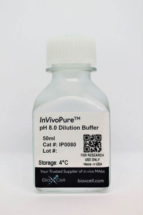
InVivoPure pH 8.0 Dilution Buffer
Product Details
InVivoPure™ dilution buffers are specifically formulated and tested to satisfy the stringent requirements for in vivo applications. They are extremely low in endotoxin, have been screened for murine pathogens, tested in animal models for toxicity and are formulated with respect to buffer composition and pH to satisfy the requirements of Bio X Cell’s antibodies.Specifications
| Endotoxin |
<0.5 EU/mL (<0.0005EU/μL) Endotoxin level is determined using an LAL gel clotting test |
|---|---|
| Sterility | 0.2 μM filtered |
| Murine Pathogen Tests |
Mouse Norovirus: Negative Mouse Parvovirus: Negative Mouse Minute Virus: Negative Mouse Hepatitis Virus: Negative Reovirus Screen: Negative Lymphocytic Choriomeningitis virus: Negative Lactate Dehydrogenase-Elevating Virus: Negative Mouse Rotavirus: Negative Theiler’s Murine Encephalomyelitis: Negative Ectromelia/Mousepox Virus: Negative Hantavirus: Negative Polyoma Virus: Negative Mouse Adenovirus: Negative Sendai Virus: Negative Mycoplasma Pulmonis: Negative Pneumonia Virus of Mice: Negative Mouse Cytomegalovirus: Negative K Virus: Negative |
| Toxicity Test Results | Nontoxic and nonantigenic in animal models |
| Concentration | 1X |
| Volume | 50 ml |
| Composition |
35 mM Na2HPO4 1.7 mM NaH2PO4 136 mM NaCl This buffer does not contain calcium, magnesium, phenol red, or preservatives such as azide. Keep contents sterile. Open only in a biological safety cabinet. |
| Storage | 4°C |
- Genetics,
IFN-γ blockade after genetic inhibition of PD-1 aggravates skeletal muscle damage and impairs skeletal muscle regeneration.
In Cellular Molecular Biology Letters on 4 April 2023 by Zhuang, S., Russell, A., et al.
PubMed
Innate immune responses play essential roles in skeletal muscle recovery after injury. Programmed cell death protein 1 (PD-1) contributes to skeletal muscle regeneration by promoting macrophage proinflammatory to anti-inflammatory phenotype transition. Interferon (IFN)-γ induces proinflammatory macrophages that appear to hinder myogenesis in vitro. Therefore, we tested the hypothesis that blocking IFN-γ in PD-1 knockout mice may dampen inflammation and promote skeletal muscle regeneration via regulating the macrophage phenotype and neutrophils. Anti-IFN-γ antibody was administered in PD-1 knockout mice, and cardiotoxin (CTX) injection was performed to induce acute skeletal muscle injury. Hematoxylin and eosin (HE) staining was used to view morphological changes of injured and regenerated skeletal muscle. Masson's trichrome staining was used to assess the degree of fibrosis. Gene expressions of proinflammatory and anti-inflammatory factors, fibrosis-related factors, and myogenic regulator factors were determined by real-time polymerase chain reaction (PCR). Changes in macrophage phenotype were examined by western blot and real-time PCR. Immunofluorescence was used to detect the accumulation of proinflammatory macrophages, anti-inflammatory macrophages, and neutrophils. IFN-γ blockade in PD-1 knockout mice did not alleviate skeletal muscle damage or improve regeneration following acute cardiotoxin-induced injury. Instead, it exacerbated skeletal muscle inflammation and fibrosis, and impaired regeneration via inhibiting macrophage accumulation, blocking macrophage proinflammatory to anti-inflammatory transition, and enhancing infiltration of neutrophils. IFN-γ is crucial for efficient skeletal muscle regeneration in the absence of PD-1. © 2023. The Author(s).

