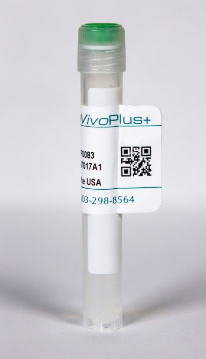
InVivoPlus mouse IgG1 isotype control, unknown specificity
Product Details
The MOPC-21 monoclonal antibody is ideal for use as a non-reactive isotype-matched control for mouse IgG1 antibodies in most in vivo and in vitro applications.Specifications
| Isotype | Mouse IgG1, κ |
|---|---|
| Recommended Dilution Buffer | InVivoPure pH 6.5 Dilution Buffer |
| Conjugation | This product is unconjugated. Conjugation is available via our Antibody Conjugation Services. |
| Formulation |
PBS, pH 6.5 Contains no stabilizers or preservatives |
| Endotoxin* |
<1EU/mg (<0.001EU/μg) Determined by LAL gel clotting assay |
| Aggregation* |
<5% Determined by SEC |
| Purity |
>95% Determined by SDS-PAGE |
| Sterility | 0.2 μM filtered |
| Production | Purified from cell culture supernatant in an animal-free facility |
| Purification | Protein G |
| RRID | AB_1107784 |
| Molecular Weight | 150 kDa |
| Murine Pathogen Tests* |
Ectromelia/Mousepox Virus: Negative Hantavirus: Negative K Virus: Negative Lactate Dehydrogenase-Elevating Virus: Negative Lymphocytic Choriomeningitis virus: Negative Mouse Adenovirus: Negative Mouse Cytomegalovirus: Negative Mouse Hepatitis Virus: Negative Mouse Minute Virus: Negative Mouse Norovirus: Negative Mouse Parvovirus: Negative Mouse Rotavirus: Negative Mycoplasma Pulmonis: Negative Pneumonia Virus of Mice: Negative Polyoma Virus: Negative Reovirus Screen: Negative Sendai Virus: Negative Theiler’s Murine Encephalomyelitis: Negative |
| Storage | The antibody solution should be stored at the stock concentration at 4°C. Do not freeze. |
Additional Formats
Recommended Products
-
Recommended Dilution Buffer
InVivoPure pH 6.5 Dilution Buffer
Faraco, G., et al. (2018). "Dietary salt promotes neurovascular and cognitive dysfunction through a gut-initiated TH17 response" Nat Neurosci 21(2): 240-249. PubMed
A diet rich in salt is linked to an increased risk of cerebrovascular diseases and dementia, but it remains unclear how dietary salt harms the brain. We report that, in mice, excess dietary salt suppresses resting cerebral blood flow and endothelial function, leading to cognitive impairment. The effect depends on expansion of TH17 cells in the small intestine, resulting in a marked increase in plasma interleukin-17 (IL-17). Circulating IL-17, in turn, promotes endothelial dysfunction and cognitive impairment by the Rho kinase-dependent inhibitory phosphorylation of endothelial nitric oxide synthase and reduced nitric oxide production in cerebral endothelial cells. The findings reveal a new gut-brain axis linking dietary habits to cognitive impairment through a gut-initiated adaptive immune response compromising brain function via circulating IL-17. Thus, the TH17 cell-IL-17 pathway is a putative target to counter the deleterious brain effects induced by dietary salt and other diseases associated with TH17 polarization.
Macal, M., et al. (2018). "Self-Renewal and Toll-like Receptor Signaling Sustain Exhausted Plasmacytoid Dendritic Cells during Chronic Viral Infection" Immunity 48(4): 730-744 e735. PubMed
Although characterization of T cell exhaustion has unlocked powerful immunotherapies, the mechanisms sustaining adaptations of short-lived innate cells to chronic inflammatory settings remain unknown. During murine chronic viral infection, we found that concerted events in bone marrow and spleen mediated by type I interferon (IFN-I) and Toll-like receptor 7 (TLR7) maintained a pool of functionally exhausted plasmacytoid dendritic cells (pDCs). In the bone marrow, IFN-I compromised the number and the developmental capacity of pDC progenitors, which generated dysfunctional pDCs. Concurrently, exhausted pDCs in the periphery were maintained by self-renewal via IFN-I- and TLR7-induced proliferation of CD4(-) subsets. On the other hand, pDC functional loss was mediated by TLR7, leading to compromised IFN-I production and resistance to secondary infection. These findings unveil the mechanisms sustaining a self-perpetuating pool of functionally exhausted pDCs and provide a framework for deciphering long-term exhaustion of other short-lived innate cells during chronic inflammation.
Manlove, L. S., et al. (2015). "Adaptive Immunity to Leukemia Is Inhibited by Cross-Reactive Induced Regulatory T Cells" J Immunol . PubMed
BCR-ABL+ acute lymphoblastic leukemia patients have transient responses to current therapies. However, the fusion of BCR to ABL generates a potential leukemia-specific Ag that could be a target for immunotherapy. We demonstrate that the immune system can limit BCR-ABL+ leukemia progression although ultimately this immune response fails. To address how BCR-ABL+ leukemia escapes immune surveillance, we developed a peptide: MHC class II tetramer that labels endogenous BCR-ABL-specific CD4+ T cells. Naive mice harbored a small population of BCR-ABL-specific T cells that proliferated modestly upon immunization. The small number of naive BCR-ABL-specific T cells was due to negative selection in the thymus, which depleted BCR-ABL-specific T cells. Consistent with this observation, we saw that BCR-ABL-specific T cells were cross-reactive with an endogenous peptide derived from ABL. Despite this cross-reactivity, the remaining population of BCR-ABL reactive T cells proliferated upon immunization with the BCR-ABL fusion peptide and adjuvant. In response to BCR-ABL+ leukemia, BCR-ABL-specific T cells proliferated and converted into regulatory T (Treg) cells, a process that was dependent on cross-reactivity with self-antigen, TGF-beta1, and MHC class II Ag presentation by leukemic cells. Treg cells were critical for leukemia progression in C57BL/6 mice, as transient Treg cell ablation led to extended survival of leukemic mice. Thus, BCR-ABL+ leukemia actively suppresses antileukemia immune responses by converting cross-reactive leukemia-specific T cells into Treg cells.
Sell, S., et al. (2015). "Control of murine cytomegalovirus infection by gammadelta T cells" PLoS Pathog 11(2): e1004481. PubMed
Infections with cytomegalovirus (CMV) can cause severe disease in immunosuppressed patients and infected newborns. Innate as well as cellular and humoral adaptive immune effector functions contribute to the control of CMV in immunocompetent individuals. None of the innate or adaptive immune functions are essential for virus control, however. Expansion of gammadelta T cells has been observed during human CMV (HCMV) infection in the fetus and in transplant patients with HCMV reactivation but the protective function of gammadelta T cells under these conditions remains unclear. Here we show for murine CMV (MCMV) infections that mice that lack CD8 and CD4 alphabeta-T cells as well as B lymphocytes can control a MCMV infection that is lethal in RAG-1(-/-) mice lacking any T- and B-cells. gammadelta T cells, isolated from infected mice can kill MCMV infected target cells in vitro and, importantly, provide long-term protection in infected RAG-1(-/-) mice after adoptive transfer. gammadelta T cells in MCMV infected hosts undergo a prominent and long-lasting phenotypic change most compatible with the view that the majority of the gammadelta T cell population persists in an effector/memory state even after resolution of the acute phase of the infection. A clonotypically focused Vgamma1 and Vgamma2 repertoire was observed at later stages of the infection in the organs where MCMV persists. These findings add gammadelta T cells as yet another protective component to the anti-CMV immune response. Our data provide clear evidence that gammadelta T cells can provide an effective control mechanism of acute CMV infections, particularly when conventional adaptive immune mechanisms are insufficient or absent, like in transplant patient or in the developing immune system in utero. The findings have implications in the stem cell transplant setting, as antigen recognition by gammadelta T cells is not MHC-restricted and dual reactivity against CMV and tumors has been described.
Leon, B., et al. (2014). "FoxP3+ regulatory T cells promote influenza-specific Tfh responses by controlling IL-2 availability" Nat Commun 5: 3495. PubMed
Here, we test the role of FoxP3(+) regulatory T cells (Tregs) in controlling T follicular helper (Tfh) and germinal centre (GC) B-cell responses to influenza. In contrast to the idea that Tregs suppress T-cell responses, we find that Treg depletion severely reduces the Tfh cell response to influenza virus. Furthermore, Treg depletion prevents the accumulation of influenza-specific GCs. These effects are not due to alterations in TGFbeta availability or a precursor-progeny relationship between Tregs and Tfh cells, but are instead mediated by increased availability of IL-2, which suppresses the differentiation of Tfh cells and as a consequence, compromises the GC B response. Thus, Tregs promote influenza-specific GC responses by preventing excessive IL-2 signalling, which suppresses Tfh cell differentiation.
Beug, S. T., et al. (2014). "Smac mimetics and innate immune stimuli synergize to promote tumor death" Nat Biotechnol 32(2): 182-190. PubMed
Smac mimetic compounds (SMC), a class of drugs that sensitize cells to apoptosis by counteracting the activity of inhibitor of apoptosis (IAP) proteins, have proven safe in phase 1 clinical trials in cancer patients. However, because SMCs act by enabling transduction of pro-apoptotic signals, SMC monotherapy may be efficacious only in the subset of patients whose tumors produce large quantities of death-inducing proteins such as inflammatory cytokines. Therefore, we reasoned that SMCs would synergize with agents that stimulate a potent yet safe “cytokine storm.” Here we show that oncolytic viruses and adjuvants such as poly(I:C) and CpG induce bystander death of cancer cells treated with SMCs that is mediated by interferon beta (IFN-beta), tumor necrosis factor alpha (TNF-alpha) and/or TNF-related apoptosis-inducing ligand (TRAIL). This combinatorial treatment resulted in tumor regression and extended survival in two mouse models of cancer. As these and other adjuvants have been proven safe in clinical trials, it may be worthwhile to explore their clinical efficacy in combination with SMCs.
Perng, O. A., et al. (2014). "The degree of CD4+ T cell autoreactivity determines cellular pathways underlying inflammatory arthritis" J Immunol 192(7): 3043-3056. PubMed
Although therapies targeting distinct cellular pathways (e.g., anticytokine versus anti-B cell therapy) have been found to be an effective strategy for at least some patients with inflammatory arthritis, the mechanisms that determine which pathways promote arthritis development are poorly understood. We have used a transgenic mouse model to examine how variations in the CD4(+) T cell response to a surrogate self-peptide can affect the cellular pathways that are required for arthritis development. CD4(+) T cells that are highly reactive with the self-peptide induce inflammatory arthritis that affects male and female mice equally. Arthritis develops by a B cell-independent mechanism, although it can be suppressed by an anti-TNF treatment, which prevented the accumulation of effector CD4(+) Th17 cells in the joints of treated mice. By contrast, arthritis develops with a significant female bias in the context of a more weakly autoreactive CD4(+) T cell response, and B cells play a prominent role in disease pathogenesis. In this setting of lower CD4(+) T cell autoreactivity, B cells promote the formation of autoreactive CD4(+) effector T cells (including Th17 cells), and IL-17 is required for arthritis development. These studies show that the degree of CD4(+) T cell reactivity for a self-peptide can play a prominent role in determining whether distinct cellular pathways can be targeted to prevent the development of inflammatory arthritis.
Vokaer, B., et al. (2013). "IL-17A and IL-2-expanded regulatory T cells cooperate to inhibit Th1-mediated rejection of MHC II disparate skin grafts" PLoS One 8(10): e76040. PubMed
Several evidences suggest that regulatory T cells (Treg) promote Th17 differentiation. Based on this hypothesis, we tested the effect of IL-17A neutralization in a model of skin transplantation in which long-term graft survival depends on a strong in vivo Treg expansion induced by transient exogenous IL-2 administration. As expected, IL-2 supplementation prevented rejection of MHC class II disparate skin allografts but, surprisingly, not in IL-17A-deficient recipients. We attested that IL-17A was not required for IL-2-mediated Treg expansion, intragraft recruitment or suppressive capacities. Instead, IL-17A prevented allograft rejection by inhibiting Th1 alloreactivity independently of Tregs. Indeed, T-bet expression of naive alloreactive CD4+ T cells and the subsequent Th1 immune response was significantly enhanced in IL-17A deficient mice. Our results illustrate for the first time a protective role of IL-17A in CD4+-mediated allograft rejection process.
Kerzerho, J., et al. (2013). "Programmed cell death ligand 2 regulates TH9 differentiation and induction of chronic airway hyperreactivity" J Allergy Clin Immunol 131(4): 1048-1057, 1057 e1041-1042. PubMed
BACKGROUND: Asthma is defined as a chronic inflammatory disease of the airways; however, the underlying physiologic and immunologic processes are not fully understood. OBJECTIVE: The aim of this study was to determine whether TH9 cells develop in vivo in a model of chronic airway hyperreactivity (AHR) and what factors control this development. METHOD: We have developed a novel chronic allergen exposure model using the clinically relevant antigen Aspergillus fumigatus to determine the time kinetics of TH9 development in vivo. RESULTS: TH9 cells were detectable in the lungs after chronic allergen exposure. The number of TH9 cells directly correlated with the severity of AHR, and anti-IL-9 treatment decreased airway inflammation. Moreover, we have identified programmed cell death ligand (PD-L) 2 as a negative regulator of TH9 cell differentiation. Lack of PD-L2 was associated with significantly increased TGF-beta and IL-1alpha levels in the lungs, enhanced pulmonary TH9 differentiation, and higher morbidity in the sensitized mice. CONCLUSION: Our findings suggest that PD-L2 plays a pivotal role in the regulation of TH9 cell development in chronic AHR, providing novel strategies for modulating adaptive immunity during chronic allergic responses.
Myles, I. A., et al. (2013). "Signaling via the IL-20 receptor inhibits cutaneous production of IL-1beta and IL-17A to promote infection with methicillin-resistant Staphylococcus aureus" Nat Immunol 14(8): 804-811. PubMed
Staphylococcus aureus causes most infections of human skin and soft tissue and is a major infectious cause of mortality. Host defense mechanisms against S. aureus are incompletely understood. Interleukin 19 (IL-19), IL-20 and IL-24 signal through type I and type II IL-20 receptors and are associated with inflammatory skin diseases such as psoriasis and atopic dermatitis. We found here that those cytokines promoted cutaneous infection with S. aureus in mice by downregulating IL-1beta- and IL-17A-dependent pathways. We noted similar effects of those cytokines in human keratinocytes after exposure to S. aureus, and antibody blockade of the IL-20 receptor improved outcomes in infected mice. Our findings identify an immunosuppressive role for IL-19, IL-20 and IL-24 during infection that could be therapeutically targeted to alter susceptibility to infection.
Lamere, M. W., et al. (2011). "Regulation of antinucleoprotein IgG by systemic vaccination and its effect on influenza virus clearance" J Virol 85(10): 5027-5035. PubMed
Seasonal influenza epidemics recur due to antigenic drift of envelope glycoprotein antigens and immune evasion of circulating viruses. Additionally, antigenic shift can lead to influenza pandemics. Thus, a universal vaccine that protects against multiple influenza virus strains could alleviate the continuing impact of this virus on human health. In mice, accelerated clearance of a new viral strain (cross-protection) can be elicited by prior infection (heterosubtypic immunity) or by immunization with the highly conserved internal nucleoprotein (NP). Both heterosubtypic immunity and NP-immune protection require antibody production. Here, we show that systemic immunization with NP readily accelerated clearance of a 2009 pandemic H1N1 influenza virus isolate in an antibody-dependent manner. However, human immunization with trivalent inactivated influenza virus vaccine (TIV) only rarely and modestly boosted existing levels of anti-NP IgG. Similar results were observed in mice, although the reaction could be enhanced with adjuvants, by adjusting the stoichiometry among NP and other vaccine components, and by increasing the interval between TIV prime and boost. Importantly, mouse heterosubtypic immunity that had waned over several months could be enhanced by injecting purified anti-NP IgG or by boosting with NP protein, correlating with a long-lived increase in anti-NP antibody titers. Thus, current immunization strategies poorly induce NP-immune antibody that is nonetheless capable of contributing to long-lived cross-protection. The high conservation of NP antigen and the known longevity of antibody responses suggest that the antiviral activity of anti-NP IgG may provide a critically needed component of a universal influenza vaccine.
Anti-S100A4 antibody administration alleviates bronchial epithelial-mesenchymal transition in asthmatic mice.
In Open Medicine (Warsaw, Poland) on 24 October 2023 by Liu, S., Liu, M., et al.
PubMed
We elucidated the effect of S100A4 on airway remodeling by regulating airway inflammation and epithelial-mesenchymal transition (EMT) in mouse models of asthma. Asthmatic mouse models were established by sensitization and challenged with ovalbumin (OVA). Anti-S100A4 antibody or control IgG antibody was administered daily before the OVA challenge. After the last challenge, airway inflammation and airway hyperresponsiveness were measured; lung tissues and bronchoalveolar lavage fluid (BALF) were harvested. Lung tissue sections were stained and evaluated for pathological changes. Levels of inflammatory cytokines were measured using ELISA. Levels of S100A4 and EMT markers were determined via western blotting analysis. Human bronchial epithelial cells were stimulated with 100 mg/mL house dust mites (HDMs) to evaluate the effect of S100A4 downregulation on EMT in vitro. S100A4 was increased in lung tissues and BALF from asthmatic mice. The asthmatic mice presented airway hyperresponsiveness, airway inflammation, and airway remodeling. After anti-S100A4 antibody administration, pathophysiological signs, including airway hyperresponsiveness and increased infiltration of inflammatory cells, were attenuated. Additionally, anti-S100A4 administration downregulated vimentin and α-SMA expression and upregulated E-cadherin expression in OVA-challenged mice. S100A4 downregulation also inhibited EMT process in HDM-stimulated 16HBE cells. Anti-S100A4 antibody administration alters airway remodeling by preventing EMT in mouse models of asthma. © 2023 the author(s), published by De Gruyter.
- Cancer Research,
TGF-β Type I Receptor Signaling in Melanoma Liver Metastases Increases Metastatic Outgrowth.
In International Journal of Molecular Sciences on 12 May 2023 by Marvin, D. L., Dijkstra, J., et al.
PubMed
Despite advances in treatment for metastatic melanoma patients, patients with liver metastasis have an unfavorable prognosis. A better understanding of the development of liver metastasis is needed. The multifunctional cytokine Transforming Growth Factor β (TGF-β) plays various roles in melanoma tumors and metastasis, affecting both tumor cells and cells from the surrounding tumor microenvironment. To study the role of TGF-β in melanoma liver metastasis, we created a model to activate or repress the TGF-β receptor pathway in vitro and in vivo in an inducible manner. For this, we engineered B16F10 melanoma cells to have inducible ectopic expression of a constitutively active (ca) or kinase-inactive (ki) TGF-β receptor I, also termed activin receptor-like kinase (ALK5). In vitro, stimulation with TGF-β signaling and ectopic caALK5 expression reduced B16F10 cell proliferation and migration. Contrasting results were found in vivo; sustained caALK5 expression in B16F10 cells in vivo increased the metastatic outgrowth in liver. Blocking microenvironmental TGF-β did not affect metastatic liver outgrowth of both control and caALK5 expressing B16F10 cells. Upon characterizing the tumor microenvironment of control and caALk5 expressing B16F10 tumors, we observed reduced (cytotoxic) T cell presence and infiltration, as well as an increase in bone marrow-derived macrophages in caALK5 expressing B16F10 tumors. This suggests that caALK5 expression in B16F10 cells induces changes in the tumor microenvironment. A comparison of newly synthesized secreted proteins upon caALK5 expression by B16F10 cells revealed increased secretion of matrix remodeling proteins. Our results show that TGF-β receptor activation in B16F10 melanoma cells can increase metastatic outgrowth in liver in vivo, possibly through remodeling of the tumor microenvironment leading to altered infiltration of immune cells. These results provide insights in the role of TGF-β signaling in B16F10 liver metastasis and could have implications regarding the use of TGF-β inhibitors for the treatment of melanoma patients with liver metastasis.
- Biochemistry and Molecular biology
Blockade of CD47 function attenuates restenosis by promoting smooth muscle cell efferocytosis and inhibiting their migration and proliferation.
In The Journal of Biological Chemistry on 1 April 2023 by Govatati, S., Pichavaram, P., et al.
PubMed
Cluster of differentiation 47 (CD47) plays an important role in the pathophysiology of various diseases including atherosclerosis but its role in neointimal hyperplasia which contributes to restenosis has not been studied. Using molecular approaches in combination with a mouse vascular endothelial denudation model, we studied the role of CD47 in injury-induced neointimal hyperplasia. We determined that thrombin-induced CD47 expression both in human aortic smooth muscle cells (HASMCs) and mouse aortic smooth muscle cells. In exploring the mechanisms, we found that the protease-activated receptor 1-Gα protein q/11 (Gαq/11)-phospholipase Cβ3-nuclear factor of activated T cells c1 signaling axis regulates thrombin-induced CD47 expression in HASMCs. Depletion of CD47 levels using its siRNA or interference of its function by its blocking antibody (bAb) blunted thrombin-induced migration and proliferation of HASMCs and mouse aortic smooth muscle cells. In addition, we found that thrombin-induced HASMC migration requires CD47 interaction with integrin β3. On the other hand, thrombin-induced HASMC proliferation was dependent on CD47's role in nuclear export and degradation of cyclin-dependent kinase-interacting protein 1. In addition, suppression of CD47 function by its bAb rescued HASMC efferocytosis from inhibition by thrombin. We also found that vascular injury induces CD47 expression in intimal SMCs and that inhibition of CD47 function by its bAb, while alleviating injury-induced inhibition of SMC efferocytosis, attenuated SMC migration, and proliferation resulting in reduced neointima formation. Thus, these findings reveal a pathological role for CD47 in neointimal hyperplasia. Copyright © 2023 The Authors. Published by Elsevier Inc. All rights reserved.
- In Vivo,
- Mus musculus (House mouse),
- Cancer Research
Tumor PD-L1 engages myeloid PD-1 to suppress type I interferon to impair cytotoxic T lymphocyte recruitment.
In Cancer Cell on 13 March 2023 by Klement, J. D., Redd, P. S., et al.
PubMed
The cellular and molecular mechanisms underlying tumor cell PD-L1 (tPD-L1) function in tumor immune evasion are incompletely understood. We report here that tPD-L1 does not suppress cytotoxic T lymphocyte (CTL) activity in co-cultures of tumor cells and tumor-specific CTLs and exhibits no effect on primary tumor growth. However, deleting tPD-L1 decreases lung metastasis in a CTL-dependent manner in tumor-bearing mice. Depletion of myeloid cells or knocking out PD-1 in myeloid cells (mPD-1) impairs tPD-L1 promotion of tumor lung metastasis in mice. Single-cell RNA sequencing (scRNA-seq) reveals that tPD-L1 engages mPD-1 to activate SHP2 to antagonize the type I interferon (IFN-I) and STAT1 pathway to repress Cxcl9 and impair CTL recruitment to lung metastases. Human cancer patient response to PD-1 blockade immunotherapy correlates with IFN-I response in myeloid cells. Our findings determine that tPD-L1 engages mPD-1 to activate SHP2 to suppress the IFN-I-STAT1-CXCL9 pathway to impair CTL tumor recruitment in lung metastasis. Copyright © 2023 The Author(s). Published by Elsevier Inc. All rights reserved.
- Cancer Research
Targeting the secreted RGDKGE collagen fragment reduces PD‑L1 by a proteasome‑dependent mechanism and inhibits tumor growth.
In Oncology Reports on 1 February 2023 by Caron, J. M., Han, X., et al.
PubMed
Structural alterations of collagen impact signaling that helps control tumor progression and the responses to therapeutic intervention. Integrins represent a class of receptors that include members that mediate collagen signaling. However, a strategy of directly targeting integrins to control tumor growth has demonstrated limited activity in the clinical setting. New molecular understanding of integrins have revealed that these receptors can regulate both pro‑ and anti‑tumorigenic functions in a cell type‑dependent manner. Therefore, designing strategies that block pro‑tumorigenic signaling, without impeding anti‑tumorigenic functions, may lead to development of more effective therapies. In the present study, evidence was provided for a novel signaling cascade in which β3‑integrin‑mediated binding to a secreted RGDKGE‑containing collagen fragment stimulates an autocrine‑like signaling pathway that differentially governs the activity of both YAP and (protein kinase‑A) PKA, ultimately leading to alterations in the levels of immune checkpoint molecule PD‑L1 by a proteasome dependent mechanism. Selectively targeting this collagen fragment, reduced nuclear YAP levels, and enhanced PKA and proteasome activity, while also exhibiting significant antitumor activity in vivo. The present findings not only provided new mechanistic insight into a previously unknown autocrine‑like signaling pathway that may provide tumor cells with the ability to regulate PD‑L1, but our findings may also help in the development of more effective strategies to control pro‑tumorigenic β3‑integrin signaling without disrupting its tumor suppressive functions in other cellular compartments.
- Immunology and Microbiology
Toxin expression during Staphylococcus aureus infection imprints host immunity to inhibit vaccine efficacy.
In NPJ Vaccines on 24 January 2023 by Teymournejad, O., Li, Z., et al.
PubMed
Staphylococcus aureus infections are a major public health issue, and a vaccine is urgently needed. Despite a considerable promise in preclinical models, all vaccines tested thus far have failed to protect humans against S. aureus. Unlike laboratory mice, humans are exposed to S. aureus throughout life. In the current study, we hypothesized that prior exposure to S. aureus "imprints" the immune response to inhibit vaccine-mediated protection. We established a mouse model in which S. aureus skin and soft tissue infection (SSTI) is followed by vaccination and secondary SSTI. Unlike naïve mice, S. aureus-sensitized mice were incompletely protected against secondary SSTI by vaccination with the inactivated α-hemolysin (Hla) mutant HlaH35L. Inhibition of protection was specific for the HlaH35L vaccine and required hla expression during primary SSTI. Surprisingly, inhibition occurred at the level of vaccine-elicited effector T cells; hla expression during primary infection limited the expansion of T cells and dendritic cells and impaired vaccine-specific T cell responses. Importantly, the T cell-stimulating adjuvant CAF01 rescued inhibition and restored vaccine-mediated protection. Together, these findings identify a potential mechanism for the failure of translation of promising S. aureus vaccines from mouse models to clinical practice and suggest a path forward to prevent these devastating infections. © 2023. The Author(s).
- Cancer Research
Expansion of interferon inducible gene pool via USP18 inhibition promotes cancer cell pyroptosis.
In Nature Communications on 17 January 2023 by Arimoto, K. I., Miyauchi, S., et al.
PubMed
While immunotherapy has emerged as a breakthrough cancer therapy, it is only effective in some patients, indicating the need of alternative therapeutic strategies. Induction of cancer immunogenic cell death (ICD) is one promising way to elicit potent adaptive immune responses against tumor-associated antigens. Type I interferon (IFN) is well known to play important roles in different aspects of immune responses, including modulating ICD in anti-tumor action. However, how to expand IFN effect in promoting ICD responses has not been addressed. Here we show that depletion of ubiquitin specific protease 18 (USP18), a negative regulator of IFN signaling, selectively induces cancer cell ICD. Lower USP18 expression correlates with better survival across human selected cancer types and delays cancer progression in mouse models. Mechanistically, nuclear USP18 controls the enhancer landscape of cancer cells and diminishes STAT2-mediated transcription complex binding to IFN-responsive elements. Consequently, USP18 suppression not only enhances expression of canonical IFN-stimulated genes (ISGs), but also activates the expression of a set of atypical ISGs and NF-κB target genes, including genes such as Polo like kinase 2 (PLK2), that induce cancer pyroptosis. These findings may support the use of targeting USP18 as a potential cancer immunotherapy. © 2023. The Author(s).
- COVID-19,
- Immunology and Microbiology
Amphiphile-CpG vaccination induces potent lymph node activation and COVID-19 immunity in mice and non-human primates.
In NPJ Vaccines on 28 October 2022 by Seenappa, L. M., Jakubowski, A., et al.
PubMed
Despite the success of currently authorized vaccines for the reduction of severe COVID-19 disease risk, rapidly emerging viral variants continue to drive pandemic waves of infection, resulting in numerous global public health challenges. Progress will depend on future advances in prophylactic vaccine activity, including advancement of candidates capable of generating more potent induction of cross-reactive T cells and durable cross-reactive antibody responses. Here we evaluated an Amphiphile (AMP) adjuvant, AMP-CpG, admixed with SARS-CoV-2 Spike receptor binding domain (RBD) immunogen, as a lymph node-targeted protein subunit vaccine (ELI-005) in mice and non-human primates (NHPs). AMP-mediated targeting of CpG DNA to draining lymph nodes resulted in comprehensive local immune activation characterized by extensive transcriptional reprogramming, inflammatory proteomic milieu, and activation of innate immune cells as key orchestrators of antigen-directed adaptive immunity. Prime-boost immunization with AMP-CpG in mice induced potent and durable T cell responses in multiple anatomical sites critical for prophylactic efficacy and prevention of severe disease. Long-lived memory responses were rapidly expanded upon re-exposure to antigen. In parallel, RBD-specific antibodies were long-lived, and exhibited cross-reactive recognition of variant RBD. AMP-CpG-adjuvanted prime-boost immunization in NHPs was safe and well tolerated, while promoting multi-cytokine-producing circulating T cell responses cross-reactive across variants of concern (VOC). Expansion of RBD-specific germinal center (GC) B cells in lymph nodes correlated to rapid seroconversion with variant-specific neutralizing antibody responses exceeding those measured in convalescent human plasma. These results demonstrate the promise of lymph-node adjuvant-targeting to coordinate innate immunity and generate robust adaptive responses critical for vaccine efficacy. © 2022. The Author(s).
Early IL-17A Prevention Rather Than Late IL-17A Neutralization Attenuates Toluene Diisocyanate-Induced Mixed Granulocytic Asthma.
In Allergy, Asthma Immunology Research on 1 September 2022 by Chen, S., Yu, L., et al.
PubMed
Interleukin (IL)-17A plays a critical role in the pathogenesis of allergic airway inflammation. Yet, the exact roles of IL-17A in asthma are still controversial. Thus, the aim of this study was to dissect the roles of IL-17A in toluene diisocyanate (TDI)-induced mixed granulocytic asthma and to assess the effects of neutralizing antibody in different effector phases on TDI-induced asthma. IL-17A functions in allergic airway inflammation were evaluated using mice deficient in IL-17A (Il17a-/-) or IL-17A monoclonal antibody (IL-17A mab, intraperitoneally, 50 μg per mouse, 100 μg per mouse). Moreover, the effects of exogenous recombinant IL (rIL)-17A in vivo (murine rIL-17A, intranasally, 1 μg per mouse) and in vitro (human rIL-17A, 100 ng/mL) were investigated. TDI-induced mixed granulocytic airway inflammation was IL-17A-dependent because airway hyperreactivity, neutrophil and eosinophil infiltration, airway smooth muscle thickness, epithelium injury, dysfunctional T helper (Th) 2 and Th17 responses, granulocytic chemokine production and mucus overproduction were more markedly reduced in the Il17a-/- mice or by IL-17A neutralization during the sensitization phase of wild-type (WT) mice. By contrast, IL-17A neutralization during the antigen-challenge phase aggravated TDI-induced eosinophils recruitment, with markedly elevated Th2 response. In line with this, instillation of rIL-17 during antigen sensitization exacerbated airway inflammation by promoting neutrophils aggregation, while rIL-17A during the antigen-challenge phase protected the mice from TDI-induced airway eosinophilia. Moreover, rIL-17A exerted distinct effects on eosinophil- or neutrophil-related signatures in vitro. Our data demonstrated that IL-17A was required for the initiation of TDI-induced asthma, but functioned as a negative regulator of established allergic inflammation, suggesting that early abrogation of IL-17A signaling, but not late IL-17A neutralization, may prevent the progression of TDI-induced asthma and could be used as a therapeutic strategy for severe asthmatics in clinical settings. Copyright © 2022 The Korean Academy of Asthma, Allergy and Clinical Immunology • The Korean Academy of Pediatric Allergy and Respiratory Disease.
- Immunology and Microbiology
SIRPα - CD47 axis regulates dendritic cell-T cell interactions and TCR activation during T cell priming in spleen.
In PLoS ONE on 13 April 2022 by Autio, A., Wang, H., et al.
PubMed
The SIRPα-CD47 axis plays an important role in T cell recruitment to sites of immune reaction and inflammation but its role in T cell antigen priming is incompletely understood. Employing OTII TCR transgenic mice bred to Cd47-/- (Cd47KO) or SKI mice, a knock-in transgenic animal expressing non-signaling cytoplasmic-truncated SIRPα, we investigated how the SIRPα-CD47 axis contributes to antigen priming. Here we show that adoptive transfer of Cd47KO or SKI Ova-specific CD4+ T cells (OTII) into Cd47KO and SKI recipients, followed by Ova immunization, elicited reduced T cell division and proliferation indices, increased apoptosis, and reduced expansion compared to transfer into WT mice. We confirmed prior reports that splenic T cell zone, CD4+ conventional dendritic cells (cDCs) and CD4+ T cell numbers were reduced in Cd47KO and SKI mice. We report that in vitro derived DCs from Cd47KO and SKI mice exhibited impaired migration in vivo and exhibited reduced CD11c+ DC proximity to OTII T cells in T cell zones after Ag immunization, which correlates with reduced TCR activation in transferred OTII T cells. These findings suggest that reduced numbers of CD4+ cDCs and their impaired migration contributes to reduced T cell-DC proximity in splenic T cell zone and reduced T cell TCR activation, cell division and proliferation, and indirectly increased T cell apoptosis.
- FC/FACS,
- Neuroscience
CD47 antibody blockade suppresses microglia-dependent phagocytosis and monocyte transition to macrophages, impairing recovery in EAE.
In JCI Insight on 8 November 2021 by Wang, H., Newton, G., et al.
PubMed
Experimental autoimmune encephalomyelitis (EAE) is a well-characterized animal model of multiple sclerosis. During the early phase of EAE, infiltrating monocytes and monocyte-derived macrophages contribute to T cell recruitment, especially CD4+ T cells, into the CNS, resulting in neuronal demyelination; however, in later stages, they promote remyelination and recovery by removal of myelin debris by phagocytosis. Signal regulatory protein α and CD47 are abundantly expressed in the CNS, and deletion of either molecule is protective in myelin oligodendrocyte glycoprotein-induced EAE because of failed effector T cell expansion and trafficking. Here we report that treatment with the function blocking CD47 Ab Miap410 substantially reduced the infiltration of pathogenic immune cells but impaired recovery from paresis. The underlying mechanism was by blocking the emergence of CD11chiMHCIIhi microglia at peak disease that expressed receptors for phagocytosis, scavenging, and lipid catabolism, which mediated clearance of myelin debris and the transition of monocytes to macrophages in the CNS. In the recovery phase of EAE, Miap410 Ab-treated mice had worsening paresis with sustained inflammation and limited remyelination as compared with control Ab-treated mice. In summary, Ab blockade of CD47 impaired resolution of CNS inflammation, thus worsening EAE.
- In Vivo,
- Mus musculus (House mouse),
- Cancer Research
Microbiota triggers STING-type I IFN-dependent monocyte reprogramming of the tumor microenvironment.
In Cell on 14 October 2021 by Lam, K. C., Araya, R. E., et al.
PubMed
The tumor microenvironment (TME) influences cancer progression and therapy response. Therefore, understanding what regulates the TME immune compartment is vital. Here we show that microbiota signals program mononuclear phagocytes in the TME toward immunostimulatory monocytes and dendritic cells (DCs). Single-cell RNA sequencing revealed that absence of microbiota skews the TME toward pro-tumorigenic macrophages. Mechanistically, we show that microbiota-derived stimulator of interferon genes (STING) agonists induce type I interferon (IFN-I) production by intratumoral monocytes to regulate macrophage polarization and natural killer (NK) cell-DC crosstalk. Microbiota modulation with a high-fiber diet triggered the intratumoral IFN-I-NK cell-DC axis and improved the efficacy of immune checkpoint blockade (ICB). We validated our findings in individuals with melanoma treated with ICB and showed that the predicted intratumoral IFN-I and immune compositional differences between responder and non-responder individuals can be transferred by fecal microbiota transplantation. Our study uncovers a mechanistic link between the microbiota and the innate TME that can be harnessed to improve cancer therapies. Published by Elsevier Inc.
- Immunology and Microbiology
Endogenous retroviruses promote homeostatic and inflammatory responses to the microbiota.
In Cell on 8 July 2021 by Lima-Júnior, D. S., Krishnamurthy, S. R., et al.
PubMed
The microbiota plays a fundamental role in regulating host immunity. However, the processes involved in the initiation and regulation of immunity to the microbiota remain largely unknown. Here, we show that the skin microbiota promotes the discrete expression of defined endogenous retroviruses (ERVs). Keratinocyte-intrinsic responses to ERVs depended on cyclic GMP-AMP synthase (cGAS)/stimulator of interferon genes protein (STING) signaling and promoted the induction of commensal-specific T cells. Inhibition of ERV reverse transcription significantly impacted these responses, resulting in impaired immunity to the microbiota and its associated tissue repair function. Conversely, a lipid-enriched diet primed the skin for heightened ERV- expression in response to commensal colonization, leading to increased immune responses and tissue inflammation. Together, our results support the idea that the host may have co-opted its endogenous virome as a means to communicate with the exogenous microbiota, resulting in a multi-kingdom dialog that controls both tissue homeostasis and inflammation.Published by Elsevier Inc.
- Immunology and Microbiology
Knockout of MAPK Phosphatase-1 Exaggerates Type I IFN Response during Systemic Escherichia coli Infection.
In The Journal of Immunology on 15 June 2021 by Kirk, S. G., Murphy, P. R., et al.
PubMed
We have previously shown that Mkp-1-deficient mice produce elevated TNF-α, IL-6, and IL-10 following systemic Escherichia coli infection, and they exhibited increased mortality, elevated bacterial burden, and profound metabolic alterations. To understand the function of Mkp-1 during bacterial infection, we performed RNA-sequencing analysis to compare the global gene expression between E. coli-infected wild-type and Mkp-1 -/- mice. A large number of IFN-stimulated genes were more robustly expressed in E. coli-infected Mkp-1 -/- mice than in wild-type mice. Multiplex analysis of the serum cytokine levels revealed profound increases in IFN-β, IFN-γ, TNF-α, IL-1α and β, IL-6, IL-10, IL-17A, IL-27, and GMSF levels in E. coli-infected Mkp-1 -/- mice relative to wild-type mice. Administration of a neutralizing Ab against the receptor for type I IFN to Mkp-1 -/- mice prior to E. coli infection augmented mortality and disease severity. Mkp-1 -/- bone marrow-derived macrophages (BMDM) produced higher levels of IFN-β mRNA and protein than did wild-type BMDM upon treatment with LPS, E. coli, polyinosinic:polycytidylic acid, and herring sperm DNA. Augmented IFN-β induction in Mkp-1 -/- BMDM was blocked by a p38 inhibitor but not by an JNK inhibitor. Enhanced Mkp-1 expression abolished IFN-β induction by both LPS and E. coli but had little effect on the IFN-β promoter activity in LPS-stimulated RAW264.7 cells. Mkp-1 deficiency did not have an overt effect on IRF3/7 phosphorylation or IKK activation but modestly enhanced IFN-β mRNA stability in LPS-stimulated BMDM. Our results suggest that Mkp-1 regulates IFN-β production primarily through a p38-mediated mechanism and that IFN-β plays a beneficial role in E. coli-induced sepsis. Copyright © 2021 by The American Association of Immunologists, Inc.
- Cancer Research
Positron Emission Tomography Imaging of Functional Transforming Growth Factor β (TGFβ) Activity and Benefit of TGFβ Inhibition in Irradiated Intracranial Tumors.
In International Journal of Radiation Oncology, Biology, Physics on 1 February 2021 by Gonzàlez-Juncà, A., Reiners, O., et al.
PubMed
Transforming growth factor β (TGFβ) promotes cell survival by endorsing DNA damage repair and mediates an immunosuppressive tumor microenvironment. Thus, TGFβ activation in response to radiation therapy is potentially targetable because it opposes therapeutic control. Strategies to assess this potential in the clinic are needed. We evaluated positron emission tomography (PET) to image 89Zr -fresolimumab, a humanized TGFβ neutralizing monoclonal antibody, as a means to detect TGFβ activation in intracranial tumor models. Pathway activity of TGFβ was validated by immunodetection of phosphorylated SMAD2 and the TGFβ target, tenascin. The contribution of TGFβ to radiation response was assessed by Kaplan-Meier survival analysis of mice bearing intracranial murine tumor models GL261 and SB28 glioblastoma and brain-adapted 4T1 breast cancer (4T1-BrA) treated with TGFβ neutralizing monoclonal antibody, 1D11, and/or focal radiation (10 Gy). 89Zr-fresolimumab PET imaging detected engineered, physiological, and radiation-induced TGFβ activation, which was confirmed by immunostaining of biological markers. GL261 glioblastoma tumors had a greater PET signal compared with similar-sized SB28 glioblastoma tumors, whereas the widespread PET signal of 4T1-BrA intracranial tumors was consistent with their highly dispersed histologic distribution. Survival of mice bearing intracranial tumors treated with 1D11 neutralizing antibody alone was similar to that of mice treated with control antibody, whereas 1D11 improved survival when given in combination with focal radiation. The extent of survival benefit of a combination of radiation and 1D11 was associated with the degree of TGFβ activity detected by PET. This study demonstrates that 89Zr-fresolimumab PET imaging detects radiation-induced TGFβ activation in tumors. Functional imaging indicated a range of TGFβ activity in intracranial tumors, but TGFβ blockade provided survival benefit only in the context of radiation treatment. This study provides further evidence that radiation-induced TGFβ activity opposes therapeutic response to radiation. Copyright © 2020 Elsevier Inc. All rights reserved.
- Cancer Research
Identification of Prostaglandin F2 Receptor Negative Regulator (PTGFRN) as an internalizable target in cancer cells for antibody-drug conjugate development.
In PLoS ONE on 28 January 2021 by Márquez, J., Dong, J., et al.
PubMed
Antibody-drug conjugates (ADC) are effective antibody-based therapeutics for hematopoietic and lymphoid tumors. However, there is need to identify new targets for ADCs, particularly for solid tumors and cancers with unmet needs. From a hybridoma library developed against cancer cells, we selected the mouse monoclonal antibody 33B7, which was able to bind to, and internalize, cancer cell lines. This antibody was used for identification of the target by immunoprecipitation and mass spectrometric analysis, followed by target validation. After target validation, 33B7 binding and target positivity were tested by flow cytometry and western blot analysis in several cancer cell lines. The ability of 33B7 conjugated to saporin to inhibit in vitro proliferation of PTFRN positive cell lines was investigated, as well as the 33B7 ADC in vivo effect on tumor growth in athymic mice. All flow cytometry and in vitro internalization assays were analyzed for statistical significance using a Welsh's T-test. Animal studies were analyzed using Two-Way Analysis of Variance (ANOVA) utilizing post-hoc Bonferroni analysis, and/or Mixed Effects analysis. The 33B7 cell surface target was identified as Prostaglandin F2 Receptor Negative Regulator (PTGFRN), a transmembrane protein in the Tetraspanin family. This target was confirmed by showing that PTGFRN-expressing cells bound and internalized 33B7, compared to PTGFRN negative cells. Cells able to bind 33B7 were PTGFRN-positive by Western blot analysis. In vitro treatment PTGFRN-positive cancer cell lines with the 33B7-saporin ADC inhibited their proliferation in a dose-dependent fashion. 33B7 conjugated to saporin was also able to block tumor growth in vivo in mouse xenografts when compared to a control ADC. These findings show that screening antibody libraries for internalizing antibodies in cancer cell lines is a good approach to identify new cancer targets for ADC development. These results suggest PTGFRN is a possible therapeutic target via antibody-based approach for certain cancers.
- Immunology and Microbiology
Human FcRn expression and Type I Interferon signaling control Echovirus 11 pathogenesis in mice.
In PLoS Pathogens on 1 January 2021 by Wells, A. I., Grimes, K. A., et al.
PubMed
Neonatal echovirus infections are characterized by severe hepatitis and neurological complications that can be fatal. Here, we show that expression of the human homologue of the neonatal Fc receptor (hFcRn), the primary receptor for echoviruses, and ablation of type I interferon (IFN) signaling are key host determinants involved in echovirus pathogenesis. We show that expression of hFcRn alone is insufficient to confer susceptibility to echovirus infections in mice. However, expression of hFcRn in mice deficient in type I interferon (IFN) signaling, hFcRn-IFNAR-/-, recapitulate the echovirus pathogenesis observed in humans. Luminex-based multianalyte profiling from E11 infected hFcRn-IFNAR-/- mice revealed a robust systemic immune response to infection, including the induction of type I IFNs. Furthermore, similar to the severe hepatitis observed in humans, E11 infection in hFcRn-IFNAR-/- mice caused profound liver damage. Our findings define the host factors involved in echovirus pathogenesis and establish in vivo models that recapitulate echovirus disease in humans.
- Immunology and Microbiology
MOSPD2 is a therapeutic target for the treatment of CNS inflammation.
In Clinical and Experimental Immunology on 1 August 2020 by Yacov, N., Kafri, P., et al.
PubMed
In multiple sclerosis and experimental autoimmune encephalomyelitis (EAE), myeloid cells comprise a major part of the inflammatory infiltrate in the central nervous system (CNS). We previously described that motile sperm domain-containing protein 2 (MOSPD2) is expressed on human myeloid cells and regulates monocyte migration in vitro. The role of MOSPD2 in EAE pathogenesis was studied by generating MOSPD2 knock-out (KO) mice and monoclonal antibodies directed against MOSPD2. We found that EAE development in MOSPD2 KO mice was significantly suppressed. While frequency representation of leukocyte subsets in lymphoid tissues was comparable, the ratio of inflammatory monocytes in the blood was markedly reduced in MOSPD2 KO mice. In addition, T cells from MOSPD2 KO mice displayed reduced secretion of proinflammatory cytokines and increased production of interleukin (IL)-4. Prophylactic and post-onset treatment using monoclonal antibodies (mAbs) generated against MOSPD2 abrogated development and reduced EAE severity. These results suggest that MOSPD2 is key in regulating migration of inflammatory monocytes, and that anti-MOSPD2 mAbs constitute a potential therapy for the treatment of CNS inflammatory diseases. © 2020 The Authors. Clinical Experimental Immunology published by John Wiley and Sons Ltd on behalf of British Society for Immunology.
- FC/FACS,
- Mus musculus (House mouse),
- Cancer Research
Proimmunogenic impact of MEK inhibition synergizes with agonist anti-CD40 immunostimulatory antibodies in tumor therapy.
In Nature Communications on 1 May 2020 by Baumann, D., Hägele, T., et al.
PubMed
Cancer types with lower mutational load and a non-permissive tumor microenvironment are intrinsically resistant to immune checkpoint blockade. While the combination of cytostatic drugs and immunostimulatory antibodies constitutes an attractive concept for overcoming this refractoriness, suppression of immune cell function by cytostatic drugs may limit therapeutic efficacy. Here we show that targeted inhibition of mitogen-activated protein kinase (MAPK) kinase (MEK) does not impair dendritic cell-mediated T cell priming and activation. Accordingly, combining MEK inhibitors (MEKi) with agonist antibodies (Abs) targeting the immunostimulatory CD40 receptor results in potent synergistic antitumor efficacy. Detailed analysis of the mechanism of action of MEKi shows that this drug exerts multiple pro-immunogenic effects, including the suppression of M2-type macrophages, myeloid derived suppressor cells and T-regulatory cells. The combination of MEK inhibition with agonist anti-CD40 Ab is therefore a promising therapeutic concept, especially for the treatment of mutant Kras-driven tumors such as pancreatic ductal adenocarcinoma.
- Pathology
Fusobacterium nucleatum facilitates ulcerative colitis through activating IL-17F signaling to NF-κB via the upregulation of CARD3 expression.
In The Journal of Pathology on 1 February 2020 by Chen, Y., Chen, Y., et al.
PubMed
Accumulating evidence links Fusobacterium nucleatum with ulcerative colitis (UC). The mechanism by which F. nucleatum promotes intestinal inflammation in UC remains poorly defined. Here, we first examined the abundance and impact of F. nucleatum on disease activity in UC tissues. Next, we isolated a strain of F. nucleatum from UC tissues and explored whether F. nucleatum aggravates the intestinal inflammatory response in vitro and in vivo. We also examined whether F. nucleatum infection involves the NF-κB or IL-17F signaling pathways. Our data showed that F. nucleatum was enriched in 51.78% of UC tissues and was correlated with the clinical course, clinical activity and refractory behavior of UC (p 0.05). Furthermore, we demonstrated that F. nucleatum promoted intestinal epithelial damage and the expression of the inflammatory cytokines IL-1β, Il-6, IL-17F and TNF-α. Mechanistically, F. nucleatum targeted caspase activation and recruitment domain 3 (CARD3) through NOD2 to activate the IL-17F/NF-κB pathway in vivo and in vitro. Thus, F. nucleatum orchestrates a molecular network involving CARD3 and IL-17F to control the UC process. Measuring and targeting F. nucleatum and its associated pathways will yield valuable insight into the prevention and treatment of UC. © 2019 Pathological Society of Great Britain and Ireland. Published by John Wiley Sons, Ltd. © 2019 Pathological Society of Great Britain and Ireland. Published by John Wiley Sons, Ltd.