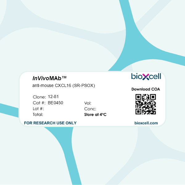
InVivoMAb anti-mouse CXCL16 (SR-PSOX)
Product Description
Specifications
| Isotype | Rat IgG1, κ |
|---|---|
| Recommended Isotype Control(s) | InVivoMAb rat IgG1 isotype control, anti-horseradish peroxidase |
| Recommended Dilution Buffer | InVivoPure pH 7.0 Dilution Buffer |
| Immunogen | mSR-PSOX-Fc fusion protein |
| Reported Applications |
in vivo neutralization of CXCL16 in vitro neutralization of CXCL16 Functional assay Flow cytometry Immunofluorescence Immunohistochemistry (frozen) |
| Formulation |
PBS, pH 7.0 Contains no stabilizers or preservatives |
| Endotoxin |
≤1EU/mg (≤0.001EU/μg) Determined by LAL assay |
| Purity |
≥95% Determined by SDS-PAGE |
| Sterility | 0.2 µm filtration |
| Production | Purified from cell culture supernatant in an animal-free facility |
| Purification | Protein G |
| Molecular Weight | 150 kDa |
| Storage | The antibody solution should be stored at the stock concentration at 4°C. Do not freeze. |
| Need a Custom Formulation? | See All Antibody Customization Options |
Application References
in vivo neutralization of CXCL16
Immunofluorescence
Zhou T, Gao Y, Wang Z, Dai C, Lei M, Liew A, Yan S, Yao Z, Hu D, Qi F (2024). "CD8 positive T-cells decrease neurogenesis and induce anxiety-like behaviour following hepatitis B vaccination" Brain Commun 6(5):fcae315.
PubMed
Mounting evidence indicates the involvement of peripheral immunity in the regulation of brain function, influencing aspects such as neuronal development, emotion, and cognitive abilities. Previous studies from our laboratory have revealed that neonatal hepatitis B vaccination can downregulate hippocampal neurogenesis, synaptic plasticity and spatial learning memory. In the current post-epidemic era characterized by universal vaccination, understanding the impact of acquired immunity on neuronal function and neuropsychiatric disorders, along with exploring potential underlying mechanisms, becomes imperative. We employed hepatitis B vaccine-induced CD3 positive T cells in immunodeficient mice to investigate the key mechanisms through which T cell subsets modulate hippocampal neurogenesis and anxiety-like behaviours. Our data revealed that mice receiving hepatitis B vaccine-induced T cells exhibited heightened anxiety and decreased hippocampal cell proliferation compared to those receiving phosphate-buffered saline-T cells or wild-type mice. Importantly, these changes were predominantly mediated by infiltrated CD8+ T cells into the brain, rather than CD4+ T cells. Transcriptome profiling of CD8+ T cells unveiled that C-X-C motif chemokine receptor 6 positive (CXCR6+) CD8+ T cells were recruited into the brain through microglial and astrocyte-derived C-X-C motif chemokine ligand 16 (CXCL16). This recruitment process impaired neurogenesis and induced anxiety-like behaviour via tumour necrosis factor-α-dependent mechanisms. Our findings highlight the role of glial cell derived CXCL16 in mediating the recruitment of CXCR6+CD8+ T cell subsets into the brain. This mechanism represents a potential avenue for modulating hippocampal neurogenesis and emotion-related behaviours after hepatitis B vaccination.
Flow Cytometry
Immunohistochemistry (frozen)
Christian LS, Wang L, Lim B, Deng D, Wu H, Wang XF, Li QJ (2021). "Resident memory T cells in tumor-distant tissues fortify against metastasis formation" Cell Rep 35(6):109118.
PubMed
As a critical machinery for rapid pathogen removal, resident memory T cells (TRMs) are locally generated after the initial encounter. However, their development accompanying tumorigenesis remains elusive. Using a murine breast cancer model, we show that TRMs develop in the tumor, the contralateral mammary mucosa, and the pre-metastatic lung. Single-cell RNA sequencing of TRMs reveals two phenotypically distinct populations representing their active versus quiescent phases. These TRMs in different tissue compartments share the same TCR clonotypes and transcriptomes with a subset of intratumoral effector/effector memory T cells (TEff/EMs), indicating their developmental ontogeny. Furthermore, CXCL16 is highly produced by tumor cells and CXCR6- TEff/EMs are the major subset preferentially egressing the tumor to form distant TRMs. Functionally, releasing CXCR6 retention in the primary tumor amplifies tumor-derived TRMs in the lung and leads to superior protection against metastases. This immunologic fortification suggests a potential strategy to prevent metastasis in clinical oncology.
in vivo neutralization of CXCL16
Wein AN, McMaster SR, Takamura S, Dunbar PR, Cartwright EK, Hayward SL, McManus DT, Shimaoka T, Ueha S, Tsukui T, Masumoto T, Kurachi M, Matsushima K, Kohlmeier JE (2019). "CXCR6 regulates localization of tissue-resident memory CD8 T cells to the air
PubMed
Resident memory T cells (TRM cells) are an important first-line defense against respiratory pathogens, but the unique contributions of lung TRM cell populations to protective immunity and the factors that govern their localization to different compartments of the lung are not well understood. Here, we show that airway and interstitial TRM cells have distinct effector functions and that CXCR6 controls the partitioning of TRM cells within the lung by recruiting CD8 TRM cells to the airways. The absence of CXCR6 significantly decreases airway CD8 TRM cells due to altered trafficking of CXCR6-/- cells within the lung, and not decreased survival in the airways. CXCL16, the ligand for CXCR6, is localized primarily at the respiratory epithelium, and mice lacking CXCL16 also had decreased CD8 TRM cells in the airways. Finally, blocking CXCL16 inhibited the steady-state maintenance of airway TRM cells. Thus, the CXCR6/CXCL16 signaling axis controls the localization of TRM cells to different compartments of the lung and maintains airway TRM cells.
Flow Cytometry
Shimaoka T, Seino K, Kume N, Minami M, Nishime C, Suematsu M, Kita T, Taniguchi M, Matsushima K, Yonehara S (2007). "Critical role for CXC chemokine ligand 16 (SR-PSOX) in Th1 response mediated by NKT cells" J Immunol 179(12):8172-9.
PubMed
The transmembrane chemokine CXCL 16 (CXCL16), which is the same molecule as the scavenger receptor that binds phosphatidylserine and oxidized lipoprotein (SR-PSOX), has been shown to mediate chemotaxis and adhesion of CXC chemokine receptor 6-expressing cells such as NKT and activated Th1 cells. We generated SR-PSOX/CXCL16-deficient mice and examined the role of this chemokine in vivo. The mutant mice showed a reduced number of liver NKT cells, and decreased production of IFN-gamma and IL-4 by administration of alpha-galactosylceramide (alphaGalCer). Of note, the alphaGalCer-induced production of IFN-gamma was more severely impaired than the production of IL-4 in SR-PSOX-deficient mice. In this context, SR-PSOX-deficient mice showed impaired sensitivity to alphaGalCer-induced anti-tumor effect mediated by IFN-gamma from NKT cells. NKT cells from wild-type mice showed impaired production of IFN-gamma, but not IL-4, after their culture with alphaGalCer and APCs from mutant mice. Moreover, Propionibacterium acnes-induced in vivo Th1 responses were severely impaired in SR-PSOX-deficient as well as NKT KO mice. Taken together, SR-PSOX/CXCL16 plays an important role in not only the production of IFN-gamma by NKT cells, but also promotion of Th1-inclined immune responses mediated by NKT cells.
in vivo neutralization of CXCL16
Hase K, Murakami T, Takatsu H, Shimaoka T, Iimura M, Hamura K, Kawano K, Ohshima S, Chihara R, Itoh K, Yonehara S, Ohno H (2006). "The membrane-bound chemokine CXCL16 expressed on follicle-associated epithelium and M cells mediates lympho-epithelial
PubMed
The recently identified CXCL16 has dual functions as a transmembrane adhesion molecule and a soluble chemokine. In this study we found that CXCL16 mRNA and protein were expressed constitutively on the follicle-associated epithelium covering Peyer's patches (PPs), isolated lymphoid follicles, and cecal patches, but minimally on the villous epithelium in the murine gastrointestinal tract. The CXCL16 receptor CXCR6/Bonzo was constitutively expressed on subpopulations of CD4+ and CD8+ T cells isolated from PPs. The expression of CXCR6/Bonzo on the PP T cells was up-regulated after stimulation with anti-CD3 and anti-CD28 mAbs. The activated PP T cells showed chemotactic migration in response to the soluble N-terminal chemokine domain of CXCL16. Furthermore, the activated PP T cells selectively adhered to cells expressing murine CXCL16. To determine the physiological role of CXCL16 in GALT, we first carefully analyzed T cell distribution in PPs. T cells localized not only in the interfollicular region but also at a lesser frequency in the subepithelial dome (SED) and in the germinal center of lymphoid follicles. Consistently, the majority of the adoptive transferred activated T cells migrated into the SED and the interfollicular region. However, the neutralization of CXCL16 specifically reduced the migration of the adoptive, transferred, activated T cells into the SED of PPs. These data suggest that CXCL16 expressed on the follicle-associated epithelium plays an important role in the recruitment and retention of activated T cells in the SED and should, at least partially, be responsible for lymphocyte compartmentalization in GALT.
in vivo neutralization of CXCL16
Nanki T, Shimaoka T, Hayashida K, Taniguchi K, Yonehara S, Miyasaka N (2005). "Pathogenic role of the CXCL16-CXCR6 pathway in rheumatoid arthritis" Arthritis Rheum 52(10):3004-14.
PubMed
Objective: Rheumatoid arthritis (RA) is a chronic inflammatory disease associated with massive T cell infiltration into the synovium. The accumulated T cells express type 1 cytokines, such as interferon-gamma (IFNgamma) and tumor necrosis factor alpha, and activated markers of inflammation, such as CD154 and inducible costimulator (ICOS). It is thought that chemokines contribute to T cell accumulation in the synovium. In this study, we examined the role of CXCL16 and CXCR6 in T cell migration and stimulation in RA synovium. Methods: Expression of CXCL16 and CXCR6 was analyzed by immunohistochemistry, reverse transcription-polymerase chain reaction, Western blotting, and/or flow cytometry. Migration activity was assessed using a chemotaxis chamber. IFNgamma production was analyzed by enzyme-linked immunosorbent assay. The effect of anti-CXCL16 monoclonal antibody on murine collagen-induced arthritis (CIA) was evaluated. Results: CXCL16 was expressed in RA synovium. CXCR6 was expressed more frequently on synovial T cells than in peripheral blood. Moreover, CXCR6-positive synovial T cells more frequently expressed CD154 and ICOS than did CXCR6-negative T cells. Stimulation with interleukin-15 (IL-15) up-regulated the expression of CXCR6 on peripheral blood T cells, and then stimulation with CXCL16 induced migration of IL-15-stimulated T cells and enhanced IFNgamma production. Furthermore, anti-CXCL16 monoclonal antibody significantly reduced the clinical arthritis score and reduced infiltration of inflammatory cells and bone destruction in the synovium of mice with CIA. Conclusion: Our results indicate that CXCL16 plays an important role in T cell accumulation and stimulation in RA synovium and suggest that CXCL16 could be a target molecule in new therapies for RA.
Flow Cytometry
Shimaoka T, Nakayama T, Fukumoto N, Kume N, Takahashi S, Yamaguchi J, Minami M, Hayashida K, Kita T, Ohsumi J, Yoshie O, Yonehara S (2004). "Cell surface-anchored SR-PSOX/CXC chemokine ligand 16 mediates firm adhesion of CXC chemokine receptor 6-expr
PubMed
Direct contacts between dendritic cells (DCs) and T cells or natural killer T (NKT) cells play important roles in primary and secondary immune responses. SR-PSOX/CXC chemokine ligand 16 (CXCL16), which is selectively expressed on DCs and macrophages, is a scavenger receptor for oxidized low-density lipoprotein and also the chemokine ligand for a G protein-coupled receptor CXC chemokine receptor 6 (CXCR6), expressed on activated T cells and NKT cells. SR-PSOX/CXCL16 is the second transmembrane-type chemokine with a chemokine domain fused to a mucin-like stalk, a structure very similar to that of fractalkine (FNK). Here, we demonstrate that SR-PSOX/CXCL16 functions as a cell adhesion molecule for cells expressing CXCR6 in the same manner that FNK functions as a cell adhesion molecule for cells expressing CX(3)C chemokine receptor 1 (CX(3)CR1) without requiring CX(3)CR1-mediated signal transduction or integrin activation. The chemokine domain of SR-PSOX/CXCL16 mediated the adhesion of CXCR6-expressing cells, which was not impaired by treatment with pertussis toxin, a Galphai protein blocker, which inhibited chemotaxis of CXCR6-expressing cells induced by SR-PSOX/CXCL16. Furthermore, the adhesion activity was up-regulated by treatment of SR-PSOX/CXCL16-expressing cells with a metalloprotease inhibitor, which increased surface expression levels of SR-PSOX/CXCL16. Thus, SR-PSOX/CXCL16 is a unique molecule that not only attracts T cells and NKT cells toward DCs but also supports their firm adhesion to DCs.
in vivo neutralization of CXCL16
in vitro neutralization of CXCL16
Functional Assays
Flow Cytometry
Fukumoto N, Shimaoka T, Fujimura H, Sakoda S, Tanaka M, Kita T, Yonehara S (2004). "Critical roles of CXC chemokine ligand 16/scavenger receptor that binds phosphatidylserine and oxidized lipoprotein in the pathogenesis of both acute and adoptive tra
PubMed
The scavenger receptor that binds phosphatidylserine and oxidized lipoprotein (SR-PSOX)/CXCL16 is a chemokine expressed on macrophages and dendritic cells, while its receptor expresses on T and NK T cells. We investigated the role of SR-PSOX/CXCL16 on acute and adoptive experimental autoimmune encephalomyelitis (EAE), which is Th1-polarized T cell-mediated autoimmune disease of the CNS. Administration of mAb against SR-PSOX/CXCL16 around the primary immunization decreased disease incidence of acute EAE with associated reduced infiltration of mononuclear cells into the CNS. Its administration was also shown to inhibit elevation of serum IFN-gamma level at primary immune response, as well as subsequent generation of Ag-specific T cells. In adoptive transfer EAE, treatment of recipient mice with anti-SR-PSOX/CXCL16 mAb also induced not only decreased clinical disease incidence, but also diminished traffic of mononuclear cells into the CNS. In addition, histopathological analyses showed that clinical development of EAE correlates well with expression of SR-PSOX/CXCL16 in the CNS. All the results show that SR-PSOX/CXCL16 plays important roles in EAE by supporting generation of Ag-specific T cells, as well as recruitment of inflammatory mononuclear cells into the CNS.

