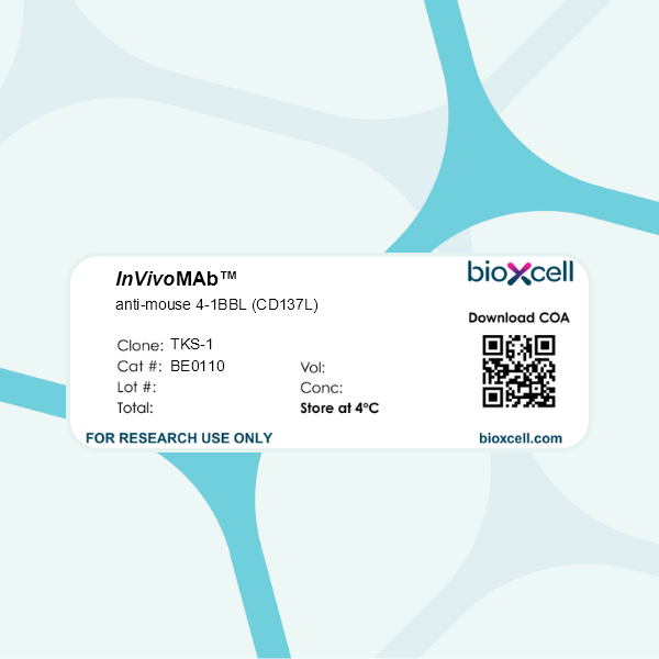
InVivoMAb anti-mouse 4-1BBL (CD137L)
Product Description
Specifications
| Isotype | Rat IgG2a, κ |
|---|---|
| Recommended Isotype Control(s) | InVivoMAb rat IgG2a isotype control, anti-trinitrophenol |
| Recommended Dilution Buffer | InVivoPure pH 7.0 Dilution Buffer |
| Conjugation | This product is unconjugated. Conjugation is available via our Antibody Conjugation Services. |
| Immunogen | Mouse 4-1BBL transfected NRK cells |
| Reported Applications |
in vivo 4-1BBL blockade ELISA |
| Formulation |
PBS, pH 7.0 Contains no stabilizers or preservatives |
| Endotoxin |
≤1EU/mg (≤0.001EU/μg) Determined by LAL assay |
| Purity |
≥95% Determined by SDS-PAGE |
| Sterility | 0.2 µm filtration |
| Production | Purified from cell culture supernatant in an animal-free facility |
| Purification | Protein G |
| RRID | AB_10949069 |
| Molecular Weight | 150 kDa |
| Storage | The antibody solution should be stored at the stock concentration at 4°C. Do not freeze. |
| Need a Custom Formulation? | See All Antibody Customization Options |
Application References
ELISA
Thomas, S., et al (2019). "Development of a new fusion-enhanced oncolytic immunotherapy platform based on herpes simplex virus type 1" J Immunother Cancer 7(1): 214.
PubMed
Oncolytic viruses preferentially replicate in tumors as compared to normal tissue and promote immunogenic cell death and induction of host systemic anti-tumor immunity. HSV-1 was chosen for further development as an oncolytic immunotherapy in this study as it is highly lytic, infects human tumor cells broadly, kills mainly by necrosis and is a potent activator of both innate and adaptive immunity. HSV-1 also has a large capacity for the insertion of additional, potentially therapeutic, exogenous genes. Finally, HSV-1 has a proven safety and efficacy profile in patients with cancer, talimogene laherparepvec (T-VEC), an oncolytic HSV-1 which expresses GM-CSF, being the only oncolytic immunotherapy approach that has received FDA approval. As the clinical efficacy of oncolytic immunotherapy has been shown to be further enhanced by combination with immune checkpoint inhibitors, developing improved oncolytic platforms which can synergize with other existing immunotherapies is a high priority. In this study we sought to further optimize HSV-1 based oncolytic immunotherapy through multiple approaches to maximize: (i) the extent of tumor cell killing, augmenting the release of tumor antigens and danger-associated molecular pattern (DAMP) factors; (ii) the immunogenicity of tumor cell death; and (iii) the resulting systemic anti-tumor immune response.
in vivo 4-1BBL blockade
Welten, S. P., et al (2015). "The viral context instructs the redundancy of costimulatory pathways in driving CD8(+) T cell expansion" Elife 4. doi : 10.7554/eLife.07486.
PubMed
Signals delivered by costimulatory molecules are implicated in driving T cell expansion. The requirements for these signals, however, vary from dispensable to essential in different infections. We examined the underlying mechanisms of this differential T cell costimulation dependence and found that the viral context determined the dependence on CD28/B7-mediated costimulation for expansion of naive and memory CD8(+) T cells, indicating that the requirement for costimulatory signals is not imprinted. Notably, related to the high-level costimulatory molecule expression induced by lymphocytic choriomeningitis virus (LCMV), CD28/B7-mediated costimulation was dispensable for accumulation of LCMV-specific CD8(+) T cells because of redundancy with the costimulatory pathways induced by TNF receptor family members (i.e., CD27, OX40, and 4-1BB). Type I IFN signaling in viral-specific CD8(+) T cells is slightly redundant with costimulatory signals. These results highlight that pathogen-specific conditions differentially and uniquely dictate the utilization of costimulatory pathways allowing shaping of effector and memory antigen-specific CD8(+) T cell responses.
in vivo 4-1BBL blockade
Kurche, J. S., et al (2010). "Comparison of OX40 ligand and CD70 in the promotion of CD4+ T cell responses" J Immunol 185(4): 2106-2115.
PubMed
The TNF superfamily members CD70 and OX40 ligand (OX40L) were reported to be important for CD4(+) T cell expansion and differentiation. However, the relative contribution of these costimulatory signals in driving CD4(+) T cell responses has not been addressed. In this study, we found that OX40L is a more important determinant than CD70 of the primary CD4(+) T cell response to multiple immunization regimens. Despite the ability of a combined TLR and CD40 agonist (TLR/CD40) stimulus to provoke appreciable expression of CD70 and OX40L on CD8(+) dendritic cells, resulting CD4(+) T cell responses were substantially reduced by Ab blockade of OX40L and, to a lesser degree, CD70. In contrast, the CD8(+) T cell responses to combined TLR/CD40 immunization were exclusively dependent on CD70. These requirements for CD4(+) and CD8(+) T cell activation were not limited to the use of combined TLR/CD40 immunization, because vaccinia virus challenge elicited primarily OX40L-dependent CD4 responses and exclusively CD70-dependent CD8(+) T cell responses. Attenuation of CD4(+) T cell priming induced by OX40L blockade was independent of signaling through the IL-12R, but it was reduced further by coblockade of CD70. Thus, costimulation by CD70 or OX40L seems to be necessary for primary CD4(+) T cell responses to multiple forms of immunization, and each may make independent contributions to CD4(+) T cell priming.
Product Citations
-
-
Immunology and Microbiology
CD137 Signaling Modulates Vein Graft Atherosclerosis by Driving T-Cell Activation and Regulating Intraplaque Angiogenesis.
In JACC Basic Transl Sci on 1 August 2025 by de Jong, A., Sluiter, T. J., et al.
PubMed
Atherosclerotic vein graft failure still presents a major problem. T-cells have been identified as one of the most abundant immune cell subset in atherosclerotic plaques. Their role, however, remains only partly understood. Using a murine vein graft model for advanced, unstable atherosclerotic lesions, we find that T-cells accumulate over time in atherosclerotic vein grafts, and appear to be activated rapidly after engraftment, demonstrated by increased expression of CD137 on plaque T-cells. Targeting of CD137 affects intraplaque angiogenesis and plaque growth, which renders CD137 a promising target for early immunomodulation to reduce atherosclerotic vein graft failure.
-
-
-
Cancer Research
-
Immunology and Microbiology
Macrophage STING signaling promotes NK cell to suppress colorectal cancer liver metastasis via 4-1BBL/4-1BB co-stimulation.
In J Immunother Cancer on 1 March 2023 by Sun, Y., Hu, H., et al.
PubMed
Macrophage innate immune response plays an important role in tumorigenesis. However, the role and mechanism of macrophage STING signaling in modulating tumor microenvironment to suppress tumor growth at secondary sites remains largely unclear.
-
-
-
Immunology and Microbiology
Activation of 4-1BBL+ B cells with CD40 agonism and IFNγ elicits potent immunity against glioblastoma.
In J Exp Med on 4 January 2021 by Lee-Chang, C., Miska, J., et al.
PubMed
Immunotherapy has revolutionized the treatment of many tumors. However, most glioblastoma (GBM) patients have not, so far, benefited from such successes. With the goal of exploring ways to boost anti-GBM immunity, we developed a B cell-based vaccine (BVax) that consists of 4-1BBL+ B cells activated with CD40 agonism and IFNγ stimulation. BVax migrates to key secondary lymphoid organs and is proficient at antigen cross-presentation, which promotes both the survival and the functionality of CD8+ T cells. A combination of radiation, BVax, and PD-L1 blockade conferred tumor eradication in 80% of treated tumor-bearing animals. This treatment elicited immunological memory that prevented the growth of new tumors upon subsequent reinjection in cured mice. GBM patient-derived BVax was successful in activating autologous CD8+ T cells; these T cells showed a strong ability to kill autologous glioma cells. Our study provides an efficient alternative to current immunotherapeutic approaches that can be readily translated to the clinic.
-
-
-
In vivo experiments
-
Mus musculus (Mouse)
Agonist-induced 4-1BB activation prevents the development of Sjӧgren's syndrome-like sialadenitis in non-obese diabetic mice.
In Biochim Biophys Acta Mol Basis Dis on 1 March 2020 by Zhou, J., You, B. R., et al.
PubMed
Activation of costimulatory receptor 4-1BB enhances T helper 1 (Th1) and CD8 T cell responses in protective immunity, and prevents or attenuates several autoimmune diseases by increasing Treg numbers and suppressing Th17 or Th2 effector response. We undertook this study to elucidate the impact of enforced 4-1BB activation on the development of Sjögren's syndrome (SS)-like sialadenitis in non-obese diabetic (NOD) model of this disease. An anti-4-1BB agnostic antibody was intraperitoneally injected to female NOD mice aged 7 weeks, prior to the disease onset that occurs around 10-11 weeks of age, 3 times weekly for 2 weeks, and the mice were analyzed for SS pathologies at age 11 weeks. The salivary flow rate was markedly higher in the anti-4-1BB-treated NOD mice compared to the IgG-treated controls. Anti-4-1BB treatment significantly reduced the leukocyte infiltration of the submandibular glands (SMGs) and the levels of serum antinuclear antibodies. Flow cytometric analysis showed that the percentages of CD4 T cells, Th17 cells and plasmacytoid dendritic cells among SMG leukocytes were markedly reduced by anti-4-1BB treatment, in conjunction with a reduction in SMG IL-23p19 mRNA levels and serum IL-17 concentrations. Although the proportion of Tregs and IL-10 mRNA levels in SMGs were not altered by 4-1BB activation, IL-10 mRNA levels in salivary gland-draining lymph nodes and serum IL-10 concentrations were both markedly increased. While anti-4-1BB treatment did not affect the amount of Th1 cells and IFNγ mRNA in the SMGs, it increased these measurables in salivary gland-draining lymph nodes. Hence, agonistic activation of 4-1BB impedes the development of SS-like sialadenitis and hyposalivation.
-
-
-
Immunology and Microbiology
Development of a new fusion-enhanced oncolytic immunotherapy platform based on herpes simplex virus type 1.
In J Immunother Cancer on 10 August 2019 by Thomas, S., Kuncheria, L., et al.
PubMed
Oncolytic viruses preferentially replicate in tumors as compared to normal tissue and promote immunogenic cell death and induction of host systemic anti-tumor immunity. HSV-1 was chosen for further development as an oncolytic immunotherapy in this study as it is highly lytic, infects human tumor cells broadly, kills mainly by necrosis and is a potent activator of both innate and adaptive immunity. HSV-1 also has a large capacity for the insertion of additional, potentially therapeutic, exogenous genes. Finally, HSV-1 has a proven safety and efficacy profile in patients with cancer, talimogene laherparepvec (T-VEC), an oncolytic HSV-1 which expresses GM-CSF, being the only oncolytic immunotherapy approach that has received FDA approval. As the clinical efficacy of oncolytic immunotherapy has been shown to be further enhanced by combination with immune checkpoint inhibitors, developing improved oncolytic platforms which can synergize with other existing immunotherapies is a high priority. In this study we sought to further optimize HSV-1 based oncolytic immunotherapy through multiple approaches to maximize: (i) the extent of tumor cell killing, augmenting the release of tumor antigens and danger-associated molecular pattern (DAMP) factors; (ii) the immunogenicity of tumor cell death; and (iii) the resulting systemic anti-tumor immune response.
-

