As the COVID-19 pandemic progresses, the need to develop new vaccines and therapeutics intensifies. Animal models of infection play important roles in such discoveries, with mice being the most widely used animal. Unfortunately, commercially available laboratory mice are not readily infected by SARS-CoV-2. To infect cells, spike proteins on the surface of SARS-CoV-2 bind to ACE2 receptors on the surface of lung cells. The problem is that SARS-CoA-2 spike proteins only bind to human ACE2 (hACE2) and not mouse ACE2.
To overcome this problem a group of researchers, led by Dr. Jincun Zhao from Guangzhou Medical University in Guangdong, China sought to create a mouse whose lung cells expressed hACE2. First, the group created an adenovirus containing the DNA sequence of hACE2. Mouse brain cells were then infected with the adenovirus in vitro which resulted in the cells expressing hACE2. These hACE2 expressing cells could be successfully infected with SARS-CoV-2.
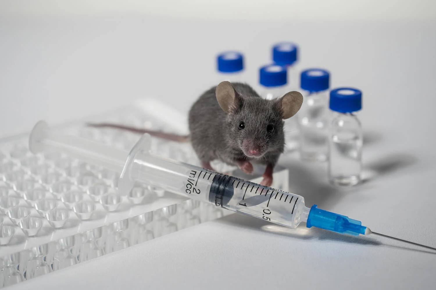
The group then infected wild-type (WT) BALB/c and C57BL/6 mice, two common laboratory strains, with the adenovirus and incredibly, these mice expressed hACE2 in their lungs. Five days later, these mice were infected with SARS-CoV-2 and within two days began to show signs of COVID-19. The mice exhibited difficulty breathing, lost up to 20% of their body weight, and had high titers of SARS-CoV-2 in their lungs. Examination of lung tissues from infected mice revealed inflammatory cell infiltrates, necrotic cell debris, and alveolar edema, all signs of severe pneumonia.Using this new COVID-19 mouse model the group discovered that type I interferon and STAT1 signaling play a critical role in viral clearance and disease resolution. The group then used Bio X Cell's anti-CD4 (clone GK1.5) and anti-CD8 (clone 2.43) antibodies to deplete CD4 and CD8 T cells in vivo in infected mice and found that these T cells were also required for optimal viral clearance.
Several therapies have been proposed to treat COVID-19, including transfer of plasma from patients who have recovered from the infection. To test this, the group obtained plasma from 3 patients who recovered from SARS-CoV-2 infection and administered the plasma to mice one day before infecting them with SARS-CoV-2. Incredibly, accelerated viral clearance and reduced lung tissue damage were observed after treatment with the plasma.
It is the hope of the authors that the availability of this COVID-19 mouse model will accelerate the pace of screening, identification, and development of drugs, vaccines, and antibody therapeutics to treat SARS-CoV-2 infection.
Read the full article in Cell.