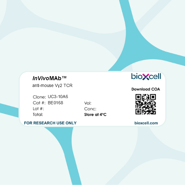
InVivoMAb anti-mouse Vγ2 TCR
Product Description
Specifications
| Isotype | Armenian Hamster IgG, κ |
|---|---|
| Recommended Isotype Control(s) | InVivoMAb polyclonal Armenian hamster IgG |
| Recommended Dilution Buffer | InVivoPure pH 7.0 Dilution Buffer |
| Conjugation | This product is unconjugated. Conjugation is available via our Antibody Conjugation Services. |
| Immunogen | G8 mouse T cells |
| Reported Applications |
in vivo γδ T cell depletion Flow cytometry |
| Formulation |
PBS, pH 7.0 Contains no stabilizers or preservatives |
| Endotoxin |
≤1EU/mg (≤0.001EU/μg) Determined by LAL assay |
| Purity |
≥95% Determined by SDS-PAGE |
| Sterility | 0.2 µm filtration |
| Production | Purified from cell culture supernatant in an animal-free facility |
| Purification | Protein G |
| RRID | AB_10950109 |
| Molecular Weight | 150 kDa |
| Storage | The antibody solution should be stored at the stock concentration at 4°C. Do not freeze. |
| Need a Custom Formulation? | See All Antibody Customization Options |
Application References
-
Rezende, R. M., et al (2018). "γδ T cells control humoral immune response by inducing T follicular helper cell differentiation" Nat Commun 9(1): 3151.
PubMed
γδ T cells have many known functions, including the regulation of antibody responses. However, how γδ T cells control humoral immunity remains elusive. Here we show that complete Freund’s adjuvant (CFA), but not alum, immunization induces a subpopulation of CXCR5-expressing γδ T cells in the draining lymph nodes. TCRγδ(+)CXCR5(+) cells present antigens to, and induce CXCR5 on, CD4 T cells by releasing Wnt ligands to initiate the T follicular helper (Tfh) cell program. Accordingly, TCRδ(-/-) mice have impaired germinal center formation, inefficient Tfh cell differentiation, and reduced serum levels of chicken ovalbumin (OVA)-specific antibodies after CFA/OVA immunization. In a mouse model of lupus, TCRδ(-/-) mice develop milder glomerulonephritis, consistent with decreased serum levels of lupus-related autoantibodies, when compared with wild type mice. Thus, modulation of the γδ T cell-dependent humoral immune response may provide a novel therapy approach for the treatment of antibody-mediated autoimmunity.
-
Hartwig, T., et al (2015). "Dermal IL-17-producing gammadelta T cells establish long-lived memory in the skin" Eur J Immunol .
PubMed
Conventional alphabeta T cells have the ability to form a long-lasting resident memory T-cell (TRM ) population in nonlymphoid tissues after encountering foreign antigen. Conversely, the concept of ‘innate memory’, where the ability of nonadaptive branches of the immune system to deliver a rapid, strengthened immune response upon reinfection or rechallenge, is just emerging. Using the alphabeta T-cell-independent Aldara psoriasis mouse model in combination with genetic fate-mapping and reporter systems, we identified a subset of gammadelta T cells in mice that is capable of establishing a long-lived memory population in the skin. IL-17A/F-producing Vgamma4+ Vdelta4+ T cells populate and persist in the dermis for long periods of time after initial stimulation with Aldara. Experienced Vgamma4+ Vdelta4+ cells show enhanced effector functions and mediate an exacerbated secondary inflammatory response. In addition to identifying a unique feature of gammadelta T cells during inflammation, our results have direct relevance to the human disease as this quasi-innate memory provides a mechanistic insight into relapses and chronification of psoriasis.
-
Suryawanshi, A., et al (2011). "Role of IL-17 and Th17 cells in herpes simplex virus-induced corneal immunopathology" J Immunol 187(4): 1919-1930.
PubMed
HSV-1 infection of the cornea leads to a blinding immunoinflammatory lesion of the eye termed stromal keratitis (SK). Recently, IL-17-producing CD4(+) T cells (Th17 cells) were shown to play a prominent role in many autoimmune conditions, but the role of IL-17 and/or of Th17 cells in virus immunopathology is unclear. In this study, we show that, after HSV infection of the cornea, IL-17 is upregulated in a biphasic manner with an initial peak production around day 2 postinfection and a second wave starting from day 7 postinfection with a steady increase until day 21 postinfection, a time point when clinical lesions are fully evident. Further studies demonstrated that innate cells, particularly gammadelta T cells, were major producers of IL-17 early after HSV infection. However, during the clinical phase of SK, the predominant source of IL-17 was Th17 cells that infiltrated the cornea only after the entry of Th1 cells. By ex vivo stimulation, the half fraction of IFN-gamma-producing CD4(+) T cells (Th1 cells) were HSV specific, whereas very few Th17 cells responded to HSV stimulation. The delayed influx of Th17 cells in the cornea was attributed to the local chemokine and cytokine milieu. Finally, HSV infection of IL-17R knockout mice as well as IL-17 neutralization in wild-type mice showed diminished SK severity. In conclusion, our results show that IL-17 and Th17 cells contribute to the pathogenesis of SK, the most common cause of infectious blindness in the Western world.
-
Martin, B., et al (2009). "Interleukin-17-producing gammadelta T cells selectively expand in response to pathogen products and environmental signals" Immunity 31(2): 321-330.
PubMed
Gammadelta T cells are an innate source of interleukin-17 (IL-17), preceding the development of the adaptive T helper 17 (Th17) cell response. Here we show that IL-17-producing T cell receptor gammadelta (TCRgammadelta) T cells share characteristic features with Th17 cells, such as expression of chemokine receptor 6 (CCR6), retinoid orphan receptor (RORgammat), aryl hydrocarbon receptor (AhR), and IL-23 receptor. AhR expression in gammadelta T cells was essential for the production of IL-22 but not for optimal IL-17 production. In contrast to Th17 cells, CCR6(+)IL-17-producing gammadelta T cells, but not other gammadelta T cells, express Toll-like receptors TLR1 and TLR2, as well as dectin-1, but not TLR4 and could directly interact with certain pathogens. This process was amplified by IL-23 and resulted in expansion, increased IL-17 production, and recruitment of neutrophils. Thus, innate receptor expression linked with IL-17 production characterizes TCRgammadelta T cells as an efficient first line of defense that can orchestrate an inflammatory response to pathogen-derived as well as environmental signals long before Th17 cells have sensed bacterial invasion.
Product Citations
-
Mammary γδ T cells promote IL-17A-mediated immunity against Staphylococcus aureus-induced mastitis in a microbiota-dependent manner.
In iScience on 15 December 2023 by Pan, N., Xiu, L., et al.
PubMed
Mastitis, a common disease for female during lactation period that could cause a health risk for human or huge economic losses for animals, is mainly caused by S. aureus invasion. Here, we found that neutrophil recruitment via IL-17A-mediated signaling was required for host defense against S. aureus-induced mastitis in a mouse model. The rapid accumulation and activation of Vγ4+ γδ T cells in the early stage of infection triggered the IL-17A-mediated immune response. Interestingly, the accumulation and influence of γδT17 cells in host defense against S. aureus-induced mastitis in a commensal microbiota-dependent manner. Overall, this study, focusing on γδT17 cells, clarified innate immune response mechanisms against S. aureus-induced mastitis, and provided a specific response to target for future immunotherapies. Meanwhile, a link between commensal microbiota community and host defense to S. aureus mammary gland infection may unveil potential therapeutic strategies to combat these intractable infections.
-
Vγ1 and Vγ4 gamma-delta T cells play opposing roles in the immunopathology of traumatic brain injury in males.
In Nat Commun on 18 July 2023 by Abou-El-Hassan, H., Rezende, R. M., et al.
PubMed
Traumatic brain injury (TBI) is a leading cause of morbidity and mortality. The innate and adaptive immune responses play an important role in the pathogenesis of TBI. Gamma-delta (γδ) T cells have been shown to affect brain immunopathology in multiple different conditions, however, their role in acute and chronic TBI is largely unknown. Here, we show that γδ T cells affect the pathophysiology of TBI as early as one day and up to one year following injury in a mouse model. TCRδ-/- mice are characterized by reduced inflammation in acute TBI and improved neurocognitive functions in chronic TBI. We find that the Vγ1 and Vγ4 γδ T cell subsets play opposing roles in TBI. Vγ4 γδ T cells infiltrate the brain and secrete IFN-γ and IL-17 that activate microglia and induce neuroinflammation. Vγ1 γδ T cells, however, secrete TGF-β that maintains microglial homeostasis and dampens TBI upon infiltrating the brain. These findings provide new insights on the role of different γδ T cell subsets after brain injury and lay down the principles for the development of targeted γδ T-cell-based therapy for TBI.
-
Vγ1 and Vγ4 γδ T cell subsets play opposing roles in traumatic brain injury
In Research Square on 2 September 2022 by Weiner, H., Abou-El-Hassan, H., et al.
-
Vγ1 and Vγ4 γδ T cell subsets play opposing roles in traumatic brain injury
In Research Square on 2 September 2022 by Weiner, H., Abou-El-Hassan, H., et al.