InVivoMAb anti-mouse TIM-3 (CD366)
Product Description
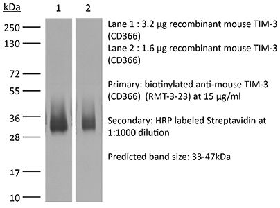
Specifications
| Isotype | Rat IgG2a, κ |
|---|---|
| Recommended Isotype Control(s) | InVivoMAb rat IgG2a isotype control, anti-trinitrophenol |
| Recommended Dilution Buffer | InVivoPure pH 7.0 Dilution Buffer |
| Conjugation | This product is unconjugated. Conjugation is available via our Antibody Conjugation Services. |
| Immunogen | Recombinant mouse TIM-3 |
| Reported Applications |
in vivo TIM-3 neutralization in vitro TIM-3 blocking Flow cytometry |
| Formulation |
PBS, pH 7.0 Contains no stabilizers or preservatives |
| Endotoxin |
≤1EU/mg (≤0.001EU/μg) Determined by LAL assay |
| Purity |
≥95% Determined by SDS-PAGE |
| Sterility | 0.2 µm filtration |
| Production | Purified from cell culture supernatant in an animal-free facility |
| Purification | Protein G |
| RRID | AB_10949464 |
| Molecular Weight | 150 kDa |
| Storage | The antibody solution should be stored at the stock concentration at 4°C. Do not freeze. |
| Need a Custom Formulation? | See All Antibody Customization Options |
Application References
-
Zelinskyy, G., et al (2011). "Virus-specific CD8+ T cells upregulate programmed death-1 expression during acute friend retrovirus infection but are highly cytotoxic and control virus replication" J Immunol 187(7): 3730-3737.
PubMed
It was recently reported that inhibitory molecules such as programmed death-1 (PD-1) were upregulated on CD8(+) T cells during acute Friend retrovirus infection and that the cells were prematurely exhausted and dysfunctional in vitro. The current study confirms that most activated CD8(+) T cells upregulated expression of PD-1 during acute infection and revealed a dichotomy of function between PD-1(hi) and PD-1(lo) subsets. More PD-1(lo) cells produced antiviral cytokines such as IFN-gamma and TNF-alpha, whereas more PD-1(hi) cells displayed characteristics of cytotoxic effectors such as production of granzymes and surface expression of CD107a. Importantly, CD8(+) T cells mediated rapid in vivo cytotoxicity and were critical for control of acute Friend virus replication. Thus, direct ex vivo analyses and in vivo experiments revealed high CD8(+) T cell functionality and indicate that PD-1 expression during acute infection is not a marker of T cell exhaustion.
-
Mittal, D., et al (2014). "Antimetastatic effects of blocking PD-1 and the adenosine A2A receptor" Cancer Res 74(14): 3652-3658.
PubMed
Adenosine targeting is an attractive new approach to cancer treatment, but no clinical study has yet examined adenosine inhibition in oncology despite the safe clinical profile of adenosine A2A receptor inhibitors (A2ARi) in Parkinson disease. Metastasis is the main cause of cancer-related deaths worldwide, and therefore we have studied experimental and spontaneous mouse models of melanoma and breast cancer metastasis to demonstrate the efficacy and mechanism of a combination of A2ARi in combination with anti-PD-1 monoclonal antibody (mAb). This combination significantly reduces metastatic burden and prolongs the life of mice compared with either monotherapy alone. Importantly, the combination was only effective when the tumor expressed high levels of CD73, suggesting a tumor biomarker that at a minimum could be used to stratify patients that might receive this combination. The mechanism of the combination therapy was critically dependent on NK cells and IFNgamma, and to a lesser extent, CD8(+) T cells and the effector molecule, perforin. Overall, these results provide a strong rationale to use A2ARi with anti-PD-1 mAb for the treatment of minimal residual and metastatic disease.
-
Dietze, K. K., et al (2013). "Combining regulatory T cell depletion and inhibitory receptor blockade improves reactivation of exhausted virus-specific CD8+ T cells and efficiently reduces chronic retroviral loads" PLoS Pathog 9(12): e1003798.
PubMed
Chronic infections with human viruses, such as HIV and HCV, or mouse viruses, such as LCMV or Friend Virus (FV), result in functional exhaustion of CD8(+) T cells. Two main mechanisms have been described that mediate this exhaustion: expression of inhibitory receptors on CD8(+) T cells and expansion of regulatory T cells (Tregs) that suppress CD8(+) T cell activity. Several studies show that blockage of one of these pathways results in reactivation of CD8(+) T cells and partial reduction in chronic viral loads. Using blocking antibodies against PD-1 ligand and Tim-3 and transgenic mice in which Tregs can be selectively ablated, we compared these two treatment strategies and combined them for the first time in a model of chronic retrovirus infection. Blocking inhibitory receptors was more efficient than transient depletion of Tregs in reactivating exhausted CD8(+) T cells and reducing viral set points. However, a combination therapy was superior to any single treatment and further augmented CD8(+) T cell responses and resulted in a sustained reduction in chronic viral loads. These results demonstrate that Tregs and inhibitory receptors are non-overlapping factors in the maintenance of chronic viral infections and that immunotherapies targeting both pathways may be a promising strategy to treat chronic infectious diseases.
-
Liu, J. F., et al (2018). "Blockade of TIM3 relieves immunosuppression through reducing regulatory T cells in head and neck cancer" J Exp Clin Cancer Res 37(1): 44.
PubMed
BACKGROUND: T-cell immunoglobulin mucin 3 (TIM3) is a negative immune checkpoint and plays a crucial part in tumor-induced immune suppression. However, the mechanism of TIM3 in regulating immunosuppression in head and neck squamous cell carcinoma (HNSCC) was still not quite clear. METHODS: We carried out the immunohistochemistry staining of HNSCC tissue microarrays. Through quantification of the histoscore, we performed the correlation analysis among the TIM3, Galectin-9, Foxp3, CD68 and CD163. The effects of TIM3 on regulatory T cells (Tregs) and macrophages were detected by utilizing the Tgfbr1/Pten 2cKO HNSCC mouse model. Flow cytometry were used to analysis the percent of Tregs, macrophages and IFN-gamma. RESULTS: We demonstrated the close association among TIM3/Galectin-9 pathway, regulatory T cell marker (Foxp3) and macrophage marker (CD68, CD163) in human HNSCC. In the transgenic HNSCC mouse model, blockade of TIM3 by the anti-TIM3 monoclonal antibody induced a reduction of CD4(+)CD25(+)Foxp3(+) Tregs. Meanwhile, the population of TIM3(+) Tregs was also decreased. However, the population of CD206(+) macrophages was not significantly declined. The increased IFN-gamma production on CD8(+) T cells in anti-TIM3 treatment mice showed that the antitumor immune response was enhanced through suppression of these negative immune factors. CONCLUSIONS: The present study demonstrated that TIM3 was associated with the immunosuppression in HNSCC. And targeting TIM3 can enhance anti-tumor immune response by decreasing Tregs in HNSCC.
Product Citations
-
Chronic inflammation-responsive hydrogel restores myeloid-T cell crosstalk to reinvigorate antitumor immunity against metastatic colorectal cancer.
In Bioact Mater on 1 August 2026 by Li, X., Fu, W., et al.
PubMed
Chronic inflammation in intermediate/advanced tumors drives burdensome protumor immune cell communication, thereby weakening immunotherapy. Conventional anti-inflammatory therapies focus on alleviating chronic inflammation whereas ignore dysfunction and scarcity of myeloid and T cells, which hinder their intercellular communication restoration. To address the dilemma, an inflammatory condition-triggered protumor inflammation-immunosurveillance shift hydrogel (TRANS) is developed to initiate adaptive immune responses mediated by intercellular communication. Triggered by inflammatory conditions, TRANS releases celecoxib (CXB) to inhibit the COX-2/PGE2 pathway, thereby reprogramming tumor-associated macrophages and mitigating protumor inflammation. Furthermore, TRANS incorporates FMS-like tyrosine kinase 3 ligands (Flt-3L) and 4-1BB agonists (α-CD137) to respectively recruit type 1 conventional DC (cDC1) and revitalize tumor-infiltrating T cells, to rejuvenate immunosurveillance. TRANS inhibits 87.50% and 88.74% of primary and secondary colorectal tumors, generates antitumor immune memory to resist tumor rechallenge, and significantly reduces lung and liver metastases. Rechallenge model shows TRANS leads to antitumor immune memory formation. Single-cell RNA sequencing is preform to elucidate the mechanism of TRANS, which exhibits that TRANS exerts antitumor effects by optimizing the crosstalk between myeloid cells and T cells via CXCL9/10-CXCR3/DPP4. TRANS further gains better control of colorectal cancer when combined with immune checkpoints inhibitors. This study offers a novel perspective on immunotherapy by rebalancing inflammation-immunity dynamics.
-
Modulating the tumor immune phenotypes by radiotherapy: formulating and validating the combination therapy of radiation, PD-L1, and TIM-3 blockade in colorectal cancer.
In J Immunother Cancer on 24 February 2026 by Wang, X. X., Zhu, C., et al.
PubMed
Most colorectal cancers (CRCs) are mismatch repair-proficient (pMMR) and microsatellite stable (MSS), and they respond poorly to immune checkpoint inhibitors (ICIs). Radiotherapy (RT) can promote antitumor immunity but may also trigger adaptive immune suppression through checkpoint upregulation, providing a rationale for combination therapies.
-
TIM-3 inhibition enhances breast tumor progression and metastasis: A paradoxical immune checkpoint response.
In J Biol Chem on 1 February 2026 by Dolui, B., Majumdar, B., et al.
PubMed
T cell immunoglobulin and mucin-domain containing-3 (TIM-3) is an emerging immune checkpoint receptor. Blocking immune checkpoint signals is a promising strategy for cancer immunotherapy. While TIM-3 blockade is currently under clinical investigation, its context-dependent role remains poorly understood. This study investigates the molecular consequences of TIM-3 inhibition using an experimental murine breast tumor model. Contrary to therapeutic expectations, administration of an anti-TIM-3 monoclonal antibody led to accelerated tumor growth and a significant increase in liver metastases. Flow cytometry revealed a paradoxical increase in tumor-infiltrating CD8+ T cells, accompanied by a reduction in CD3+ and Foxp3+ T cells. Cytokine profiling showed elevated levels of IFN-γ, TNF-α, and IL-17 in the serum, with increased IL-10 and IL-1β in tumors and altered cytokine expression in the spleen. Proteomic analysis identified 1371 dysregulated proteins, and gene set enrichment analysis revealed upregulation of PI3K/Akt-mTORC signaling, which promotes CDK4-mediated proliferation and tumor stemness via B2M and CD44. Gene ontology analysis indicated suppression of autophagy and apoptosis pathways, including downregulation of the proinflammatory protein complex calprotectin (S100A8/A9). Notably, TIM-3 blockades enhanced epithelial-to-mesenchymal transition (EMT) and c-MYC signaling, potentially driven by Foxp3 downregulation. Proteomics data are available via ProteomeXchange with identifier PXD065028. This finding challenges the prevailing view that immune checkpoint blockade uniformly suppresses tumor growth. Instead, this study demonstrates that TIM-3 inhibition may paradoxically exacerbate tumor progression and metastasis.
-
Identification of anti-TIM-3 based checkpoint inhibitor combinations with activity in immunotherapy refractory melanoma models.
In J Immunother Cancer on 18 August 2025 by Phadke, M. S., Li, J., et al.
PubMed
A significant percentage of melanomas are refractory to immune checkpoint inhibitor (ICI) monotherapies and combinations. As there are currently no effective second-line therapies available for ICI-resistant patients, we sought to identify novel checkpoint inhibitor combinations for future clinical evaluation.