
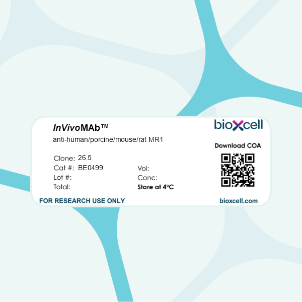
InVivoMAb anti-human/porcine/mouse/rat MR1
in vitro functional assay
Immunoprecipitation
Immunofluorescence
Flow cytometry
ELISA
Product Description
Specifications
| Isotype | Mouse IgG2a, κ |
|---|---|
| Recommended Isotype Control(s) | InVivoMAb rat IgG2a isotype control, anti-trinitrophenol |
| Recommended Dilution Buffer | InVivoPure pH 7.0 Dilution Buffer |
| Immunogen | Soluble ectodomain of insect-expressed human MR1/β2m complexes |
| Reported Applications |
in vitro blocking of MR1-dependent MAIT cell activation in vitro functional assay Immunoprecipitation Immunofluorescence Flow cytometry ELISA |
| Formulation |
PBS, pH 7.0 Contains no stabilizers or preservatives |
| Endotoxin |
≤1EU/mg (≤0.001EU/μg) Determined by LAL assay |
| Purity |
≥95% Determined by SDS-PAGE |
| Sterility | 0.2 µm filtration |
| Production | Purified from cell culture supernatant in an animal-free facility |
| Purification | Protein G |
| Molecular Weight | 150 kDa |
| Storage | This product should be stored at the stock concentration at 4°C. Do not freeze. |
| Need a Custom Formulation? | See All Antibody Customization Options |
Application References
-
Huang S, Gilfillan S, Cella M, Miley MJ, Lantz O, Lybarger L, Fremont DH, Hansen TH (2005). "Evidence for MR1 antigen presentation to mucosal-associated invariant T cells." J Biol Chem 280(22):21183-93.
PubMed
The novel class Ib molecule MR1 is highly conserved in mammals, particularly in its alpha1/alpha2 domains. Recent studies demonstrated that MR1 expression is required for development and expansion of a small population of T cells expressing an invariant T cell receptor (TCR) alpha chain called mucosal-associated invariant T (MAIT) cells. Despite these intriguing properties it has been difficult to determine whether MR1 expression and MAIT cell recognition is ligand-dependent. To address these outstanding questions, monoclonal antibodies were produced in MR1 knock-out mice immunized with recombinant MR1 protein, and a series of MR1 mutations were generated at sites previously shown to disrupt the ability of class Ia molecules to bind peptide or TCR. Here we show that 1) MR1 molecules are detected by monoclonal antibodies in either an open or folded conformation that correlates precisely with peptide-induced conformational changes in class Ia molecules, 2) only the folded MR1 conformer activated 2/2 MAIT hybridoma cells tested, 3) the pattern of MAIT cell activation by the MR1 mutants implies the MR1/TCR orientation is strikingly similar to published major histocompatibility complex/alphabetaTCR engagements, 4) all the MR1 mutations tested and found to severely reduce surface expression of folded molecules were located in the putative ligand binding groove, and 5) certain groove mutants of MR1 that are highly expressed on the cell surface disrupt MAIT cell activation. These combined data strongly support the conclusion that MR1 has an antigen presentation function.
-
Kjer-Nielsen L, Patel O, Corbett AJ, Le Nours J, Meehan B, Liu L, Bhati M, Chen Z, Kostenko L, Reantragoon R, Williamson NA, Purcell AW, Dudek NL, McConville MJ, O'Hair RA, Khairallah GN, Godfrey DI, Fairlie DP, Rossjohn J, McCluskey J (2012). "MR1 p
PubMed
Antigen-presenting molecules, encoded by the major histocompatibility complex (MHC) and CD1 family, bind peptide- and lipid-based antigens, respectively, for recognition by T cells. Mucosal-associated invariant T (MAIT) cells are an abundant population of innate-like T cells in humans that are activated by an antigen(s) bound to the MHC class I-like molecule MR1. Although the identity of MR1-restricted antigen(s) is unknown, it is present in numerous bacteria and yeast. Here we show that the structure and chemistry within the antigen-binding cleft of MR1 is distinct from the MHC and CD1 families. MR1 is ideally suited to bind ligands originating from vitamin metabolites. The structure of MR1 in complex with 6-formyl pterin, a folic acid (vitamin B9) metabolite, shows the pterin ring sequestered within MR1. Furthermore, we characterize related MR1-restricted vitamin derivatives, originating from the bacterial riboflavin (vitamin B2) biosynthetic pathway, which specifically and potently activate MAIT cells. Accordingly, we show that metabolites of vitamin B represent a class of antigen that are presented by MR1 for MAIT-cell immunosurveillance. As many vitamin biosynthetic pathways are unique to bacteria and yeast, our data suggest that MAIT cells use these metabolites to detect microbial infection.
-
Huang S, Martin E, Kim S, Yu L, Soudais C, Fremont DH, Lantz O, Hansen TH (2009). "MR1 antigen presentation to mucosal-associated invariant T cells was highly conserved in evolution." Proc Natl Acad Sci U S A 106(20):8290-5.
PubMed
Several nonclassical major histocompatibilty antigens (class Ib molecules) have emerged as key players in the early immune response to pathogens or stress. Class Ib molecules activate subsets of T cells that mount effector responses before the adaptive immune system, and thus are called innate T cells. MR1 is a novel class Ib molecule with properties highly suggestive of its regulation of mucosal immunity. The Mr1 gene is evolutionarily conserved, is non-Mhc linked, and controls the development of mucosal-associated invariant T (MAIT) cells. MAIT cells preferentially reside in the gut, and their development is dependent on commensal microbiota. Although these properties suggest that MAIT cells function as innate T cells in the mucosa, this has been difficult to test, due to the (i) paucity of MAIT cells that display MR1-specific activation in vitro and (ii) lack of knowledge of whether or not MR1 presents antigen. Here we show that both mouse and human MAIT cells display a high level of cross-reactivity on mammalian MR1 orthologs, but with differences consistent with limited ligand discrimination. Furthermore, acid eluates from recombinant or cellular MR1 proteins enhance MAIT cell activation in an MR1-specific and cross-species manner. Our findings demonstrate that the presentation pathway of MR1 to MAIT cells is highly evolutionarily conserved.
-
Leeansyah E, Ganesh A, Quigley MF, Sönnerborg A, Andersson J, Hunt PW, Somsouk M, Deeks SG, Martin JN, Moll M, Shacklett BL, Sandberg JK (2013). "Activation, exhaustion, and persistent decline of the antimicrobial MR1-restricted MAIT-cell population
PubMed
Mucosal-associated invariant T (MAIT) cells are an evolutionarily conserved antimicrobial MR1-restricted T-cell subset. MAIT cells are CD161(+), express a V7.2 TCR, are primarily CD8(+) and numerous in blood and mucosal tissues. However, their role in HIV-1 infection is unknown. In this study, we found levels of MAIT cells to be severely reduced in circulation in patients with chronic HIV-1 infection. Residual MAIT cells were highly activated and functionally exhausted. Their decline was associated with time since diagnosis, activation levels, and the concomitant expansion of a subset of functionally impaired CD161(+) V7.2(+) T cells. Such cells were generated in vitro by exposure of MAIT cells to Escherichia coli. Notably, whereas the function of residual MAIT cells was at least partly restored by effective antiretroviral therapy, levels of MAIT cells in peripheral blood were not restored. Interestingly, MAIT cells in rectal mucosa were relatively preserved, although some of the changes seen in blood were recapitulated in the mucosa. These findings are consistent with a model in which the MAIT-cell compartment, possibly as a result of persistent exposure to microbial material, is engaged, activated, exhausted, and progressively and persistently depleted during chronic HIV-1 infection.