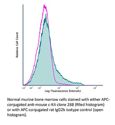
FlowMAb APC anti-mouse c-Kit (CD117)
Product Description
Specifications
| Isotype | Rat IgG2b, κ |
|---|---|
| Recommended Isotype Control(s) | FlowMAb APC rat IgG2b isotype control, anti-keyhole limpet hemocyanin |
| Conjugation | APC |
| Excitation Source | Red 627-640 nm |
| Excitation Max | 651 nm |
| Emission Max | 660 nm |
| Immunogen | Mouse bone marrow mast cells |
| Reported Applications |
Flow cytometry Immunofluorescence Immunohistochemistry |
| Protocol Information | It is recommended that the reagent be carefully titrated for optimal performance in the assay of interest. |
| Concentration | 0.2 mg/ml |
| Formulation |
PBS, pH 7.0 Contains 0.09% Sodium Azide |
| Production | Purified from cell culture supernatant in an animal-free facility |
| Purification | Protein G. Conjugated with allophycocyanin under optimal conditions. |
| Storage | The antibody solution should be stored at the stock concentration at 4°C and protected from prolonged exposure to light. Do not freeze. |
| Need a Custom Formulation? | See All Antibody Customization Options |
Application References
Flow Cytometry
Siracusa, M. C., et al. (2013). "Thymic stromal lymphopoietin-mediated extramedullary hematopoiesis promotes allergic inflammation" Immunity 39(6): 1158-1170.
PubMed
Extramedullary hematopoiesis (EMH) refers to the differentiation of hematopoietic stem cells (HSCs) into effector cells that occurs in compartments outside of the bone marrow. Previous studies linked pattern-recognition receptor (PRR)-expressing HSCs, EMH, and immune responses to microbial stimuli. However, whether EMH operates in broader immune contexts remains unknown. Here, we demonstrate a previously unrecognized role for thymic stromal lymphopoietin (TSLP) in promoting the population expansion of progenitor cells in the periphery and identify that TSLP-elicited progenitors differentiated into effector cells including macrophages, dendritic cells, and granulocytes and that these cells contributed to type 2 cytokine responses. The frequency of circulating progenitor cells was also increased in allergic patients with a gain-of-function polymorphism in TSLP, suggesting the TSLP-EMH pathway might operate in human disease. These data identify that TSLP-induced EMH contributes to the development of allergic inflammation and indicate that EMH is a conserved mechanism of innate immunity.
Flow Cytometry
Krishnamoorthy, N., et al. (2008). "Activation of c-Kit in dendritic cells regulates T helper cell differentiation and allergic asthma" Nat Med 14(5): 565-573.
PubMed
Dendritic cells (DCs) are integral to the differentiation of T helper cells into T helper type 1 T(H)1, T(H)2 and T(H)17 subsets. Interleukin-6 (IL-6) plays an important part in regulating these three arms of the immune response by limiting the T(H)1 response and promoting the T(H)2 and T(H)17 responses. In this study, we investigated pathways in DCs that promote IL-6 production. We show that the allergen house dust mite (HDM) or the mucosal adjuvant cholera toxin promotes cell surface expression of c-Kit and its ligand, stem cell factor (SCF), on DCs. This dual upregulation of c-Kit and SCF results in sustained signaling downstream of c-Kit, promoting IL-6 secretion. Intranasal administration of antigen into c-Kit-mutant mice or neutralization of IL-6 in cultures established from the lung-draining lymph nodes of immunized wild-type mice blunted the T(H)2 and T(H)17 responses. DCs lacking functional c-Kit or those unable to express membrane-bound SCF secreted lower amounts of IL-6 in response to HDM or cholera toxin. DCs expressing nonfunctional c-Kit were unable to induce a robust T(H)2 or T(H)17 response and elicited diminished allergic airway inflammation when adoptively transferred into mice. Expression of the Notch ligand Jagged-2, which has been associated with T(H)2 differentiation, was blunted in DCs from c-Kit-mutant mice. c-Kit upregulation was specifically induced by T(H)2- and T(H)17-skewing stimuli, as the T(H)1-inducing adjuvant, CpG oligodeoxynucleotide, did not promote either c-Kit or Jagged-2 expression. DCs generated from mice expressing a catalytically inactive form of the p110delta subunit of phosphatidylinositol-3 (PI3) kinase (p110(D910A)) secreted lower amounts of IL-6 upon stimulation with cholera toxin. Collectively, these results highlight the importance of the c-Kit-PI3 kinase-IL-6 signaling axis in DCs in regulating T cell responses.
Immunofluorescence
Lee, E. J., et al. (2005). "Pituitary transcription factor-1 induces transient differentiation of adult hepatic stem cells into prolactin-producing cells in vivo" Mol Endocrinol 19(4): 964-971.
PubMed
A subset of transcription factors function as pivotal regulators of cell differentiation pathways. Pituitary transcription factor-1 (Pit-1) is a tissue-specific homeodomain protein that specifies the development of pituitary somatotropes and lactotropes. In this study, adenovirus was used to deliver rat Pit-1 to mouse liver. Pit-1 expression was detected in the majority (50-80%) of hepatocyte nuclei after tail vein injection (2 x 10(9) plaque forming units). Pit-1 activated hepatic expression of the endogenous prolactin (PRL), GH, and TSHbeta genes along with several other markers of lactotrope progenitor cells. Focal clusters (0.2-0.5% of liver cells per tissue section) of periportal cells were positive for PRL by immunofluorescent staining. The PRL-producing cells also expressed proliferating cell nuclear antigen as well as the hepatic stem cell markers (c-Kit, Thy1, and cytokeratin 14). These data indicate that Pit-1 induces the transient differentiation of hepatic progenitor cells into PRL-producing cells, providing additional evidence that transcription factors can specify the differentiation pathway of adult stem cells.
Flow Cytometry
Immunohistochemistry
Li, Z., et al. (2005). "Developmental stage-selective effect of somatically mutated leukemogenic transcription factor GATA1" Nat Genet 37(6): 613-619.
PubMed
Acquired mutations in the hematopoietic transcription factor GATA binding protein-1 (GATA1) are found in megakaryoblasts from nearly all individuals with Down syndrome with transient myeloproliferative disorder (TMD, also called transient leukemia) and the related acute megakaryoblastic leukemia (DS-AMKL, also called DS-AML M7). These mutations lead to production of a variant GATA1 protein (GATA1s) that is truncated at its N terminus. To understand the biological properties of GATA1s and its relation to DS-AMKL and TMD, we used gene targeting to generate Gata1 alleles that express GATA1s in mice. We show that the dominant action of GATA1s leads to hyperproliferation of a unique, previously unrecognized yolk sac and fetal liver progenitor, which we propose accounts for the transient nature of TMD and the restriction of DS-AMKL to infants. Our observations raise the possibility that the target cells in other leukemias of infancy and early childhood are distinct from those in adult leukemias and underscore the interplay between specific oncoproteins and potential target cells.
Flow Cytometry
Chen, C. C., et al. (2005). "Identification of mast cell progenitors in adult mice" Proc Natl Acad Sci U S A 102(32): 11408-11413.
PubMed
It is well known that mast cells are derived from hematopoietic stem cells. However, in adult hematopoiesis, a committed mast cell progenitor has not yet been identified in any species, nor is it clear at what point during adult hematopoiesis commitment to the mast cell lineage occurs. We identified a cell population in adult mouse bone marrow, characterized as Lin(-)c-Kit(+)Sca-1(-)-Ly6c(-)FcepsilonRIalpha(-)CD27(-)beta7(+)T1/ST2+, that gives rise only to mast cells in culture and that can reconstitute the mast cell compartment when transferred into c-kit mutant mast cell-deficient mice. In addition, our experiments strongly suggest that these adult mast cell progenitors are derived directly from multipotential progenitors instead of, as previously proposed, common myeloid progenitors or granulocyte/macrophage progenitors.
Flow Cytometry
Ikuta, K. and I. L. Weissman. (1992). "Evidence that hematopoietic stem cells express mouse c-kit but do not depend on steel factor for their generation" Proc Natl Acad Sci U S A 89(4): 1502-1506.
PubMed
The interaction of the mouse c-kit receptor, designated Kit receptor, and steel factor promotes the proliferation and differentiation of hematopoietic progenitor cells. Monoclonal antibodies against the extracellular portion of the mouse Kit receptor were established. Five percent to 10% of total bone marrow cells expressed the Kit receptor, and half of them lack the expression of lineage markers. The Kit receptor was expressed on 70-80% of Thy-1.1lo Lin-Sca-1+ cells, which express Thy-1.1 antigen at a low level and constitute approximately 0.05% of adult bone marrow and fetal liver; by previous studies, these cells have been shown to be highly enriched for multipotent hematopoietic stem cells (HSCs) and are the only hematopoietic cell subset with this activity. Spleen colony formation and long-term multilineage reconstitution activities were contained in the Kit+ but not in the Kit- subpopulations of Thy-1lo Lin-Sca-1+ cells from adult bone marrow, suggesting that the Kit receptor is expressed on HSCs from the earliest stage-i.e., pluripotent HSCs. The role of steel factor in the development and self-renewal of HSCs was tested with Sl/Sl homozygote fetuses, which lack genes to encode functional steel factor. They were shown to have 30-40% of the number of HSCs on days 13-15 when compared with normal litermates. However, the absolute number of HSCs increased during fetal development in the Sl/Sl mice. The results suggest that the Kit receptor-steel factor interaction may not be essential for the initiation of hematopoiesis and the self-renewal of (at least) fetal HSCs.

