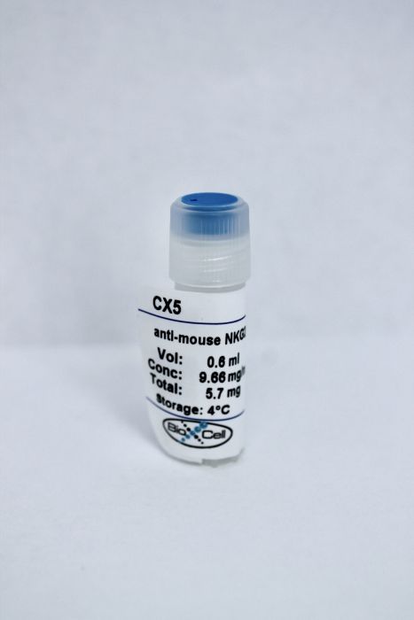
InVivoMAb anti-mouse NKG2D (CD314)
Product Details
The CX5 monoclonal antibody reacts with mouse NKG2D, a type II transmembrane lectin-like glycoprotein also known as CD314. NKG2D is expressed on NK cells, NKT cells, CD8 T cells, γ/δ T cells, and macrophages. NKG2D has been implicated in anti-tumor surveillance, the immune response against viral infection, and in diabetes progression in NOD mice. Previous studies have shown that CX5 is a non-depleting antibody, which blocks binding of NKG2D to its ligands and mediates internalization of the receptor.Specifications
| Isotype | Rat IgG1, κ |
|---|---|
| Recommended Isotype Control(s) | InVivoMAb rat IgG1 isotype control, anti-horseradish peroxidase |
| Recommended Dilution Buffer | InVivoPure pH 7.0 Dilution Buffer |
| Conjugation | This product is unconjugated. Conjugation is available via our Antibody Conjugation Services. |
| Immunogen | Purified mouse NKG2D |
| Reported Applications |
in vivo NKG2D blockade in vitro NKG2D blockade Flow Cytometry |
| Formulation |
PBS, pH 7.0 Contains no stabilizers or preservatives |
| Endotoxin |
<2EU/mg (<0.002EU/μg) Determined by LAL gel clotting assay |
| Purity |
>95% Determined by SDS-PAGE |
| Sterility | 0.2 μM filtered |
| Production | Purified from cell culture supernatant in an animal-free facility |
| Purification | Protein G |
| RRID | AB_2894754 |
| Molecular Weight | 150 kDa |
| Storage | The antibody solution should be stored at the stock concentration at 4°C. Do not freeze. |
Recommended Products
-
Recommended Isotype Control(s)
InVivoMAb rat IgG1 isotype control, anti-horseradish peroxidase
-

Recommended Dilution Buffer
InVivoPure pH 7.0 Dilution Buffer
in vivo NKG2D blockade, in vitro NKG2D blockade
Wang, D., et al. (2018). "NK cells inhibit anti-Mycobacterium bovis BCG T cell responses and aggravate pulmonary inflammation in a direct lung infection mouse model" Cell Microbiol 20(7): e12833. PubMed
Tuberculosis remains a threat to public health. The major problem for curing this disease is latent infection, of which the underlying mechanisms are still not fully understood. Previous studies indicate that natural killer (NK) cells do not play a role in inhibiting the growth of Mycobacterium tuberculosis in the lung, and recent studies have revealed that NK cells regulate the adaptive immunity during mycobacterial infection. By using a mouse model of direct lung infection with Mycobacterium bovis bacillus Calmette-Guerin (BCG), we found that the presence of NK cells postponed the priming and activation of T cells after BCG infection. In addition, depletion of NK cells before infection alleviated pulmonary pathology. Further studies showed that NK cells lysed BCG-infected macrophages in an NKG2D dependent manner. Thus, NK cells did not play a direct role in control BCG, but aggravated the pulmonary inflammation and impaired anti-BCG T cell immunity, likely through killing BCG-infected macrophages. Our results may have important implications for the design of immune therapy to treat tuberculosis.
in vivo NKG2D blockade, in vitro NKG2D blockade, Flow Cytometry
Hosomi, S., et al. (2017). "Intestinal epithelial cell endoplasmic reticulum stress promotes MULT1 up-regulation and NKG2D-mediated inflammation" J Exp Med 214(10): 2985-2997. PubMed
Endoplasmic reticulum (ER) stress is commonly observed in intestinal epithelial cells (IECs) and can, if excessive, cause spontaneous intestinal inflammation as shown by mice with IEC-specific deletion of X-box-binding protein 1 (Xbp1), an unfolded protein response-related transcription factor. In this study, Xbp1 deletion in the epithelium (Xbp1(DeltaIEC) ) is shown to cause increased expression of natural killer group 2 member D (NKG2D) ligand (NKG2DL) mouse UL16-binding protein (ULBP)-like transcript 1 and its human orthologue cytomegalovirus ULBP via ER stress-related transcription factor C/EBP homology protein. Increased NKG2DL expression on mouse IECs is associated with increased numbers of intraepithelial NKG2D-expressing group 1 innate lymphoid cells (ILCs; NK cells or ILC1). Blockade of NKG2D suppresses cytolysis against ER-stressed epithelial cells in vitro and spontaneous enteritis in vivo. Pharmacological depletion of NK1.1(+) cells also significantly improved enteritis, whereas enteritis was not ameliorated in Recombinase activating gene 1(-/-);Xbp1(DeltaIEC) mice. These experiments reveal innate immune sensing of ER stress in IECs as an important mechanism of intestinal inflammation.
in vivo NKG2D blockade, in vitro NKG2D blockade
Yang, D., et al. (2017). "NKG2D(+)CD4(+) T Cells Kill Regulatory T Cells in a NKG2D-NKG2D Ligand- Dependent Manner in Systemic Lupus Erythematosus" Sci Rep 7(1): 1288. PubMed
Systemic lupus erythematosus (SLE) features a decreased pool of CD4(+)CD25(+)Foxp3(+) T regulatory (Treg) cells. We had previously observed NKG2D(+)CD4(+) T cell expansion in contrast to a decreased pool of Treg cells in SLE patients, but whether NKG2D(+)CD4(+) T cells contribute to the decreased Treg cells remains unclear. In the present study, we found that the NKG2D(+)CD4(+) T cells efficiently killed NKG2D ligand (NKG2DL)(+) Treg cells in vitro, whereby the surviving Treg cells in SLE patients showed no detectable expression of NKG2DLs. It was further found that MRL/lpr lupus mice have significantly increased percentage of NKG2D(+)CD4(+) T cells and obvious decreased percentage of Treg cells, as compared with wild-type mice. Adoptively transferred NKG2DL(+) Treg cells were found to be efficiently killed in MRL/lpr lupus mice, with NKG2D neutralization remarkably attenuating this killing. Anti-NKG2D or anti-interferon-alpha receptor (IFNAR) antibodies treatment in MRL/lpr mice restored Treg cells numbers and markedly ameliorated the lupus disease. These results suggest that NKG2D(+)CD4(+) T cells are involved in the pathogenesis of SLE by killing Treg cells in a NKG2D-NKG2DL-dependent manner. Targeting the NKG2D-NKG2DL interaction might be a potential therapeutic strategy by which Treg cells can be protected from cytolysis in SLE patients.
in vivo NKG2D blockade
Kjellev, S., et al. (2007). "Inhibition of NKG2D receptor function by antibody therapy attenuates transfer-induced colitis in SCID mice" Eur J Immunol 37(5): 1397-1406. PubMed
A role for the activating NK-receptor NKG2D has been indicated in several autoimmune diseases in humans and in animal models of type 1 diabetes and multiple sclerosis, and treatment with monoclonal antibodies to NKG2D attenuated disease severity in these models. In an adoptive transfer-induced model of colitis, we found a significantly higher frequency of CD4(+)NKG2D(+) cells in blood, mesenteric lymph nodes, colon, and spleen from colitic mice compared to BALB/c donor-mice. We, therefore, wanted to study the effect of anti-NKG2D antibody (CX5) treatment initiated either before onset of colitis, when the colitis was mild, or when severe colitis was established. CX5 treatment decreased the detectable levels of cell-surface NKG2D and prophylactic administration of CX5 attenuated the development of colitis significantly, whereas a more moderate reduction in the severity of disease was observed after CX5 administration to mildly colitic animals. CX5 did not attenuate severe colitis. We conclude that the frequency of CD4(+)NKG2D(+) cells increase during development of experimental colitis. NKG2D may play a role in the early stages of colitis in this model, since early administration of CX5 attenuated disease severity.
in vivo NKG2D blockade, in vitro NKG2D blockade
Ogasawara, K., et al. (2004). "NKG2D blockade prevents autoimmune diabetes in NOD mice" Immunity 20(6): 757-767. PubMed
NKG2D is an activating receptor on CD8(+) T cells and NK cells that has been implicated in immunity against tumors and microbial pathogens. Here we show that RAE-1 is present in prediabetic pancreas islets of NOD mice and that autoreactive CD8(+) T cells infiltrating the pancreas express NKG2D. Treatment with a nondepleting anti-NKG2D monoclonal antibody (mAb) during the prediabetic stage completely prevented disease by impairing the expansion and function of autoreactive CD8(+) T cells. These findings demonstrate that NKG2D is essential for disease progression and suggest a new therapeutic target for autoimmune type I diabetes.