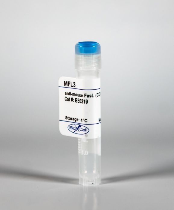
InVivoMAb anti-mouse FasL (CD178)
Product Details
The MFL3 monoclonal antibody reacts with mouse Fas Ligand (FasL) also known as CD178, CD95 Ligand, and TNFSF6. FasL is a 40 kDa type II transmembrane glycoprotein and a member of the TNF superfamily. FasL is expressed on activated T cells and in spleen, testis, and eye. Upon binding to its receptor CD95 (Fas) FasL induces apoptotic cell death to maintain peripheral tolerance. Some tumors over-express FasL and induce the apoptosis of infiltrating lymphocytes, allowing the tumor to escape the effects of an immune response. CD178/CD95 interactions are also thought to play a role in the proliferation of CD8+ cells and neutrophil extravasation, chemotaxis and survival. The MFL3 antibody has been reported to block CD178/CD95 induced apoptosis.Specifications
| Isotype | Armenian hamster IgG |
|---|---|
| Recommended Isotype Control(s) | InVivoMAb polyclonal Armenian hamster IgG |
| Recommended Dilution Buffer | InVivoPure pH 7.0 Dilution Buffer |
| Conjugation | This product is unconjugated. Conjugation is available via our Antibody Conjugation Services. |
| Immunogen | BHK cells expressing B6 mouse FasL |
| Reported Applications |
in vivo FasL blockade In vitro FasL blockade Functional assay Immunohistochemistry (paraffin) Flow Cytometry |
| Formulation |
PBS, pH 7.0 Contains no stabilizers or preservatives |
| Endotoxin |
<2EU/mg (<0.002EU/μg) Determined by LAL gel clotting assay |
| Purity |
>95% Determined by SDS-PAGE |
| Sterility | 0.2 μM filtered |
| Production | Purified from cell culture supernatant in an animal-free facility |
| Purification | Protein A |
| RRID | AB_2819046 |
| Molecular Weight | 150 kDa |
| Storage | The antibody solution should be stored at the stock concentration at 4°C. Do not freeze. |
Recommended Products
-
Recommended Isotype Control(s)
InVivoMAb polyclonal Armenian hamster IgG
-

Recommended Dilution Buffer
InVivoPure pH 7.0 Dilution Buffer
in vivo FasL blockade
Upadhyay, R., et al. (2021). "A Critical Role for Fas-Mediated Off-Target Tumor Killing in T-cell Immunotherapy" Cancer Discov 11(3): 599-613. PubMed
T cell-based therapies have induced cancer remissions, though most tumors ultimately progress, reflecting inherent or acquired resistance including antigen escape. Better understanding of how T cells eliminate tumors will help decipher resistance mechanisms. We used a CRISPR/Cas9 screen and identified a necessary role for Fas-FasL in antigen-specific T-cell killing. We also found that Fas-FasL mediated off-target “bystander” killing of antigen-negative tumor cells. This localized bystander cytotoxicity enhanced clearance of antigen-heterogeneous tumors in vivo, a finding that has not been shown previously. Fas-mediated on-target and bystander killing was reproduced in chimeric antigen receptor (CAR-T) and bispecific antibody T-cell models and was augmented by inhibiting regulators of Fas signaling. Tumoral FAS expression alone predicted survival of CAR-T-treated patients in a large clinical trial (NCT02348216). These data suggest strategies to prevent immune escape by targeting both the antigen expression of most tumor cells and the geography of antigen-loss variants. SIGNIFICANCE: This study demonstrates the first report of in vivo Fas-dependent bystander killing of antigen-negative tumors by T cells, a phenomenon that may be contributing to the high response rates of antigen-directed immunotherapies despite tumoral heterogeneity. Small molecules that target the Fas pathway may potentiate this mechanism to prevent cancer relapse.
in vivo FasL blockade
Lakins, M. A., et al. (2018). "Cancer-associated fibroblasts induce antigen-specific deletion of CD8 (+) T Cells to protect tumour cells" Nat Commun 9(1): 948. PubMed
Tumours have developed strategies to interfere with most steps required for anti-tumour immune responses. Although many populations contribute to anti-tumour responses, tumour-infiltrating cytotoxic T cells dominate, hence, many suppressive strategies act to inhibit these. Tumour-associated T cells are frequently restricted to stromal zones rather than tumour islands, raising the possibility that the tumour microenvironment, where crosstalk between malignant and “normal” stromal cells exists, may be critical for T cell suppression. We provide evidence of direct interactions between stroma and T cells driving suppression, showing that cancer-associated fibroblasts (CAFs) sample, process and cross-present antigen, killing CD8(+) T cells in an antigen-specific, antigen-dependent manner via PD-L2 and FASL. Inhibitory ligand expression is observed in CAFs from human tumours, and neutralisation of PD-L2 or FASL reactivates T cell cytotoxic capacity in vitro and in vivo. Thus, CAFs support T cell suppression within the tumour microenvironment by a mechanism dependent on immune checkpoint activation.
in vivo FasL blockade
Lotti, R., et al. (2018). "Soluble Fas Ligand Is Essential for Blister Formation in Pemphigus" Front Immunol 9: 370. PubMed
Pemphigus is a blistering disease characterized by pemphigus autoantibodies (PVIgG) directed mostly against desmogleins (Dsgs), resulting in the loss of keratinocyte adhesion (acantholysis). Yet, the mechanisms underlying blister formation remain to be clarified. We have shown previously that anti-Fas ligand (FasL) antibody (Ab) prevents PVIgG-induced caspase-8 activation and Dsg cleavage in human keratinocytes, and that sera from pemphigus patients contain abnormally increased levels of FasL. Here, we demonstrate that recombinant FasL induces the activation of caspases prior to Dsg degradation, and anti-FasL Ab prevents acantholysis in cultured keratinocytes. Moreover, the silencing of FasL reduces PVIgG-induced caspase-8 activation and Dsg3 cleavage. Following injection of PVIgG into mice, FasL is upregulated at 1-3 h and is followed by caspase-8-mediated keratinocyte apoptosis, before blister formation. The administration of anti-FasL Ab after PVIgG injection blocks blister formation in mice. Furthermore, we injected PVIgG into two different gene-targeted mutant mice that selectively lack either secreted soluble FasL (sFasL), FasL(Deltas/Deltas) mice, or the membrane-bound form of FasL (mFasL), FasL(Deltam/Deltam) mice. After PVIgG treatment, blisters are only visible in FasL(Deltam/Deltam) animals, lacking mFasL, but still producing sFasL, similar to wild-type (C57BL/6) animals. By contrast, a significant decrease in the relative acantholytic area is observed in the FasL(Deltas/Deltas) animals. These results demonstrate that soluble FasL plays a crucial role in the mechanisms of blister formation, and blockade of FasL could be an effective therapeutic approach for pemphigus.
Immunohistochemistry (paraffin)
Bien, K., et al. (2017). "Involvement of Fas/FasL pathway in the murine model of atopic dermatitis" Inflamm Res 66(8): 679-690. PubMed
OBJECTIVE AND DESIGN: The aim of this study was to elucidate the role of apoptosis mediated through Fas/FasL pathway using the mouse model of atopic dermatitis (AD). MATERIALS AND TREATMENT: AD was induced by epicutaneous application of ovalbumin (OVA) in wild-type C57BL/6, B6. MRL-Faslpr/J (Fas-) and B6Smn.C3-Faslgld/J (FasL-) mouse strains. METHODS: Skin samples were subjected to staining for Fas/FasL expression, M30 epitope and assessment of inflammatory response via immunohistochemical staining. Cytokine and chemokine production was assessed by real-time PCR. RESULTS: In comparison to wild-type mice, OVA sensitization of Fas- and FasL-deficient mice led to increased epidermal and dermal thickness, collagen deposition and local inflammation consisting of macrophages, neutrophils and CD4+ T cells. Fas- and FasL-deficient mice showed increased total counts of regulatory T cells (Tregs) and IgE levels in blood as well as increased expression of IL-1beta, IL-4, IL-5, IL-13 and TGF-1beta mRNA in comparison to wild-type mice. On the other hand, expression of CXCL9 and CXCL10, IL-17 mRNAs in the skin samples in Fas- and FasL-deficient mice was decreased. CONCLUSIONS: Our results show that lack of the Fas-induced apoptosis leads to exacerbation of AD characteristics such as Th2 inflammation and dermal thickening. Therefore, Fas receptor can play an important role in AD pathogenesis by controlling development of the local inflammation.
in vitro FasL blockade
D’Aloia, M. M., et al. (2016). "T lymphocytes engineered to express a CD16-chimeric antigen receptor redirect T-cell immune responses against immunoglobulin G-opsonized target cells" Cytotherapy 18(2): 278-290. PubMed
BACKGROUND AIMS: Chimeric antigen receptors (CARs) designed for adoptive immunotherapy need to achieve two functions: antigen recognition and triggering of the lytic machinery of reprogrammed effector cells. Cytotoxic T cells have been engineered with FcgammaRIII (CD16) chimeric molecules to be redirected against malignant cells by monoclonal antibodies (mAbs). These cells have been proven to mediate granule-dependent cellular cytotoxicity, but it is not clear whether they can also kill malignant cells by a granule-independent mechanism of cell cytotoxicity. METHODS: We engineered a CD16A-CAR equipped with the extracellular CD16A, the hinge spacer and the transmembrane region of CD8, and the zeta-chain of the T-cell receptor/CD3 complex in tandem with the CD28 co-stimulatory signal transducer module. The CD16A-CAR was expressed and functionally tested in the MD45 cell line, a murine T-cell hybridoma with a defective granular exocytosis pathway but capable of killing target cells by a Fas ligand-mediated lysis. RESULTS: Our results indicate that in vitro cross-linking of CD16A-CAR on MD45 cells by the Fc fragment of mAb opsonized tumor cells induced interleukin-2 release and granule-independent cellular cytotoxicity. CONCLUSIONS: We conclude that strategies aimed to implement the therapeutic functions of mAbs used in the clinic with T-dependent immune responses driven by engineered T cells expressing FcgammaR-CAR can boost the antitumor efficacy of mAbs used in the clinic.
in vivo FasL blockade
Linkermann, A., et al. (2011). "Renal tubular Fas ligand mediates fratricide in cisplatin-induced acute kidney failure" Kidney Int 79(2): 169-178. PubMed
Cisplatin, a standard chemotherapeutic agent for many tumors, has an unfortunately common toxicity where almost a third of patients develop renal dysfunction after a single dose. Acute kidney injury caused by cisplatin depends on Fas-mediated apoptosis driven by Fas ligand (FasL) expressed on tubular epithelial and infiltrating immune cells. Since the role of FasL in T cells is known, we investigated whether its presence in primary kidney cells is needed for its toxic effect. We found that all cisplatin-treated wild-type (wt) mice died within 6 days; however, severe combined immunodeficiency (SCID)/beige mice (B-, T-, and natural killer-cell-deficient) displayed a significant survival benefit, with only 55% mortality while exhibiting significant renal failure. Treating SCID/beige mice with MFL3, a FasL-blocking monoclonal antibody, completely restored survival after an otherwise lethal cisplatin dose, suggesting another source of FasL besides immune cells. Freshly isolated primary tubule segments from wt mice were co-incubated with thick ascending limb (TAL) segments freshly isolated from mice expressing the green fluorescent protein (GFP) transgene (same genetic background) to determine whether FasL-mediated killing of tubular cells is an autocrine or paracrine mechanism. Cisplatin-stimulated primary segments induced apoptosis in the GFP-tagged TAL cells, an effect blocked by MFL3. Thus, our study shows that cisplatin-induced nephropathy is mediated through FasL, functionally expressed on tubular cells that are capable of inducing death of cells of adjacent tubules.
Flow Cytometry, Functional Assays
Kayagaki, N., et al. (1997). "Polymorphism of murine Fas ligand that affects the biological activity" Proc Natl Acad Sci U S A 94(8): 3914-3919. PubMed
Ala-184 and Glu-218 –> Gly-218) in the extracellular region. Analysis of the K10 reactivity and genotyping by PCR-restriction fragment length polymorphism revealed that inbred mice segregate into the following two allotypes: mFasL.1 (B6, C3H, MRL, SJL, NOD, NZB, NZW) and mFasL.2 (BALB/c, DBA/1, DBA/2). Interestingly, COS7 cells expressing BALB/c FasL lysed Fas-bearing target cells more efficiently than those expressing B6 FasL. Furthermore, BALB/c-derived CD8-FasL fusion protein, which is composed of the extracellular domains of human CD8alpha and mFasL, exhibited 9-fold higher specific activity than did B6-derived CD8-FasL. These results suggest that in mFasL.2 mice the Fas/FasL system works more effectively than in mFasL.1 mice.”}” data-sheets-userformat=”{“2″:14851,”3”:{“1″:0},”4”:{“1″:2,”2″:16777215},”12″:0,”14”:{“1″:2,”2″:1521491},”15″:”Roboto, sans-serif”,”16″:12}”>Fas ligand (FasL) is a member of the tumor necrosis factor family and induces apoptosis in Fas (CD95)-bearing target cells. In this study, we generated several mAbs that react with mouse FasL (mFasL) and characterized their functional properties. One of these mAbs, K10, specifically reacted with mFasL derived from C57BL/6 (B6) mice, but not that from BALB/c mice as estimated by surface staining and blocking of cytotoxic activities of mFasL transfectants, suggesting a polymorphism of mFasL. Sequence analysis of mFasL cDNA from several strains revealed that BALB/c and DBA/2 mice have three nucleotide differences from the known B6 and C3H sequences, which result in two amino acid substitutions (Thr-184 –> Ala-184 and Glu-218 –> Gly-218) in the extracellular region. Analysis of the K10 reactivity and genotyping by PCR-restriction fragment length polymorphism revealed that inbred mice segregate into the following two allotypes: mFasL.1 (B6, C3H, MRL, SJL, NOD, NZB, NZW) and mFasL.2 (BALB/c, DBA/1, DBA/2). Interestingly, COS7 cells expressing BALB/c FasL lysed Fas-bearing target cells more efficiently than those expressing B6 FasL. Furthermore, BALB/c-derived CD8-FasL fusion protein, which is composed of the extracellular domains of human CD8alpha and mFasL, exhibited 9-fold higher specific activity than did B6-derived CD8-FasL. These results suggest that in mFasL.2 mice the Fas/FasL system works more effectively than in mFasL.1 mice.